+91 8691888178 / 8850999397 dr.jyotibansode.01@gmail.com Monday to Saturday : 05:00PM TO 06:00PM

Protein in Urine Causes: What’s Normal, What’s Worrisome?

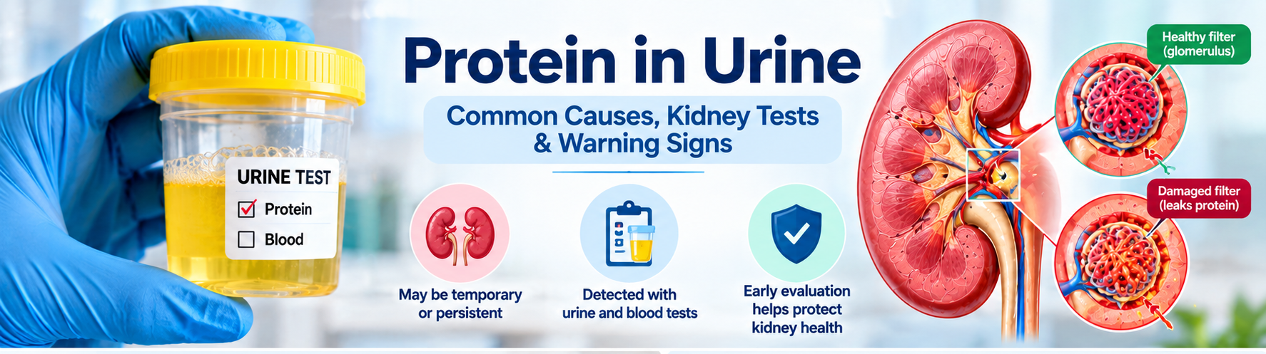
Short answer : Protein in urine can be a fleeting finding after fever, intense exercise, or dehydration, but persistent proteinuria often signals damage to the kidney’s filtering units (glomeruli). Ongoing leakage may require further testing and specialist care. Proteinuria—often discovered on a routine urine test—doesn’t always mean serious disease. A single abnormal result may be temporary, yet repeated or high‑level protein loss usually points to kidney‑filter damage that needs evaluation. What Is Proteinuria? Healthy kidneys keep large proteins such as albumin inside the bloodstream while allowing waste and excess fluid to pass into urine. The glomeruli act as tiny sieves; when they are inflamed, scarred, or otherwise damaged, proteins slip through and appear in the urine. How the kidneys normally work  – Blood enters the glomerulus at high pressure.  – Small molecules (water, electrolytes, waste) filter out. – Large proteins are retained and re‑absorbed in the tubules. When the glomerular barrier is compromised, albumin and other proteins are lost, leading to a measurable proteinuria. Common Causes of Protein in Urine (Persistent) These conditions often produce ongoing protein leakage and merit a thorough work‑up:  – Diabetes mellitus – high blood sugar damages glomerular capillaries.  – Hypertension (high blood pressure) – pressure overload injures the filtration barrier.  – Glomerular diseases – e.g., IgA nephropathy, membranous nephropathy, focal segmental glomerulosclerosis.  – Chronic infections – hepatitis C, HIV, or recurrent urinary‑tract infections.  – Cardiovascular conditions – heart failure can raise kidney venous pressure.  – Medications – non‑steroidal anti‑inflammatory drugs (NSAIDs), certain antibiotics, and some chemotherapy agents.  – Other kidney disorders – polycystic kidney disease, obstructive uropathy. For an in‑depth look at these factors, see Causes of Protein in Urine: Understanding the Key Factors. Temporary Causes of Protein in Urine Not every positive protein test signals chronic disease. Transient proteinuria often resolves once the trigger disappears: Fever or acute illness – inflammation raises glomerular permeability. Strenuous exercise – muscle breakdown and increased blood flow can spill protein. Dehydration – concentrated urine may falsely appear protein‑rich. Cold exposure – vasoconstriction can temporarily affect filtration. Recent urinary‑tract infection – inflammation of the bladder or urethra. Because a single abnormal result may be fleeting, clinicians often repeat a morning urine sample before ordering extensive work‑up. Symptoms and Warning Signs Early proteinuria is frequently silent, but watch for these clues:  – Foamy, frothy, or bubbly urine.  – Swelling (edema) of ankles, feet, or around the eyes.  – Unexplained fatigue or loss of appetite.  – Changes in urine volume or frequency.  – Elevated blood pressure. Most people with mild proteinuria feel fine; the presence of symptoms often indicates a higher level of protein loss or concurrent kidney dysfunction. Does foamy urine always mean protein in urine? No. Foam can result from rapid urine flow, concentrated urine, or even certain detergents in the toilet bowl. Only a laboratory test (dipstick or ACR) can confirm proteinuria. Protein in Urine vs. Blood in Urine Proteinuria (protein) and hematuria (blood) are distinct findings. Protein indicates a leak of albumin or larger proteins, while blood signals red‑cell leakage, often from a different part of the urinary tract. When both appear together, clinicians consider glomerular disease or severe inflammation. Read more about the overlap in our protein and blood in urine article. How Proteinuria Is Measured The most common tests include: – Urine dipstick – quick screening, semi‑quantitative.  – Urine albumin‑to‑creatinine ratio (UACR) – expresses albumin in mg per gram of creatinine; preferred for diabetes monitoring.  – Urine protein‑to‑creatinine ratio (PCR) – measures total protein.  – Comprehensive urinalysis – evaluates sediment, cells, and casts.  – Serum creatinine & eGFR – assess overall kidney filtration function. Interpretation of UACR:Range (mg/g)Interpretation<30Normal or low‑grade30–300Micro‑albuminuria (early kidney damage)>300Macro‑albuminuria (significant kidney disease) Urine ACR (UACR) Result Interpretation UACR Result Interpretation Below 30 mg/g Normal to mildly increased 30–300 mg/g Moderately increased albuminuria Above 300 mg/g Severely increased albuminuria Results should be repeated, especially if the first sample was a random collection, and interpreted alongside creatinine, eGFR, and clinical context. Can You Have Protein in Urine with Normal Creatinine? Yes. Protein leakage can precede a rise in serum creatinine because creatinine reflects overall filtration, not the selective loss of albumin. Early glomerular injury may show a high UACR while eGFR remains >90 mL/min/1.73 m². Glomerular Disease: When to Be Concerned Persistent, significant proteinuria—especially when accompanied by any of the following—should raise suspicion for a primary glomerular disorder:  – Protein levels >300 mg/g (macro‑albuminuria).  – Concurrent hematuria (blood in urine).  – Unexplained edema or high blood pressure.  – Declining eGFR over months.  – Abnormal urine sediment (casts, dysmorphic RBCs). Dr. Jyoti Bansode has a special clinical interest in glomerular diseases; patients with these red flags are often referred for nephrology evaluation. When Might a Kidney Biopsy Be Needed? Most proteinuria cases are managed without invasive procedures. A biopsy is considered when:  – The underlying cause remains unclear after standard labs and imaging.  – Protein loss exceeds 3.5 g/day (nephrotic‑range) with rapid eGFR decline.  – There is suspicion of a specific glomerular disease that guides therapy (e.g., lupus nephritis, IgA nephropathy).  – Patients have refractory proteinuria despite optimal blood‑pressure and diabetes control. Read more about the procedure in our kidney biopsy overview. Patient Checklist After an Abnormal Urine Protein Test Ask which test was performed (dipstick, UACR, PCR). Confirm the exact protein value and its units. Request your serum creatinine and eGFR results. Discuss any recent fever, intense exercise, dehydration, or infection that could be temporary. Inquire whether a repeat morning urine sample is recommended. Review blood‑pressure control and diabetes management. Ask if a referral to a nephrologist is appropriate. When to See a Nephrologist Consider specialist care if you have any of the following:  – Persistent proteinuria on two separate tests (≥30 mg/g).  – Rising UACR or PCR over weeks to months.  – Proteinuria accompanied by hematuria.  – Declining eGFR (<60 mL/min/1.73 m²) or rising serum creatinine.  – Significant edema or uncontrolled hypertension.  – Suspected glomerular disease based on urine sediment.  – Unclear cause after standard work‑up. Schedule a consultation through our appointment and consultation page. Medical ReviewDr. Jyoti
Kidney Specialist in Vashi: What to Bring to Your Consultation

Visiting a kidney specialist (nephrologist) for the first time can feel overwhelming, especially if you’re unsure which documents or test results to carry. In Vashi, NewEra Hospitals hosts Dr. Jyoti Bansode, a seasoned nephrologist with more than a decade of experience. This guide walks you through the exact items you should prepare, the symptoms that prompt a visit, and how the consultation typically unfolds, so you can feel confident and ready. When Should You See a Kidney Specialist in Vashi? Kidney health often goes unnoticed until a problem becomes evident through lab values or symptoms. According to When Should You See a Nephrologist in Navi Mumbai?, you should consider a referral when any of the following occur: Persistent swelling in the ankles, feet, or face. Unexplained fatigue or weakness despite adequate rest. Changes in urine colour, frequency, or the presence of blood. High blood pressure that is difficult to control with medication. Elevated serum creatinine or a declining eGFR on routine blood work. These red‑flag signs often indicate that a detailed renal evaluation is needed. Common Symptoms That Prompt a Nephrology Referral While some kidney issues are detected incidentally, many patients notice physical cues. Typical symptoms include: Foamy or bubbly urine – a sign of proteinuria. Persistent lower‑back or flank pain, which may suggest stones or infection. Frequent urination at night (nocturia). Unexplained itching, especially on the arms and legs. Loss of appetite or nausea. If you experience any of these, schedule an appointment promptly. Nephrology consultation checklist showing lab reports, kidney ultrasound, medications, medical history and questions to ask What to Bring to Your Consultation Arriving prepared saves time and ensures the nephrologist can make an accurate assessment. Below is a comprehensive checklist. Essential Lab Reports Blood and urine tests form the backbone of kidney evaluation. Bring the most recent reports, ideally from the past 3 months: Serum Creatinine & eGFR – shows current filtration rate. Blood Urea Nitrogen (BUN) – complements creatinine. Electrolytes (Na+, K+, Cl-, HCO3-) – reveals imbalances. Urine Albumin‑to‑Creatinine Ratio (UACR) or 24‑hour urine protein – quantifies protein loss. Lipid profile – important for cardiovascular risk in CKD patients. These tests are highlighted in What to Expect at Your First Visit with a Nephrologist, which notes that most doctors request a full panel before the first appointment. Imaging and Other Diagnostic Documents If you have already undergone imaging, bring copies of the reports: Renal ultrasound – assesses kidney size and structural abnormalities. CT or MRI scans – useful for complex stone disease or tumors. Previous kidney biopsies – include pathology results if available. Even if the imaging was performed at another facility, a printed PDF or CD will help Dr. Bansode review the findings without delay. Personal Health History Prepare a concise summary of your medical background: List of current medications, including over‑the‑counter supplements. History of diabetes, hypertension, or autoimmune diseases. Family history of kidney disease or transplant. Any prior surgeries or hospitalizations related to the urinary tract. Having this information on hand enables the doctor to spot patterns and tailor treatment. How Dr. Jyoti Bansode Evaluates Your Case Dr. Jyoti BansodeMBBS, MD Medicine, DM Nephrology10+ years of nephrology experiencePractice at NewEra Hospitals, Vashi Dr. Bansode follows a systematic approach that blends clinical expertise with evidence‑based guidelines. Review of Tests and Imaging During the appointment, the nephrologist will first examine your lab values and imaging reports. Patterns such as a steadily falling eGFR or persistent proteinuria guide the next steps—whether it’s a medication adjustment, a repeat test, or a referral for a kidney biopsy. Physical Examination The doctor will assess blood pressure (both sitting and standing), check for edema, and listen for heart or lung sounds that may indicate fluid overload. A focused abdominal exam helps identify kidney enlargement or tenderness. Discussion & Plan After the assessment, Dr. Bansode will explain the diagnosis in plain language, outline lifestyle modifications, and prescribe any necessary medications. She also discusses the need for follow‑up labs, potential referrals (e.g., dietitian, transplant team), and answers any questions you have. Checklist for Your Appointment What to bring to a nephrology consultation including kidney test reports, imaging, medications and medical history Symptoms to Mention: swelling, foamy urine, nocturia, fatigue, itching. Lab Reports: serum creatinine, eGFR, BUN, electrolytes, urine protein, lipid profile. Imaging: renal ultrasound, CT/MRI, biopsy reports (if any). Medication List: include doses, frequency, and any herbal supplements. Health History: diabetes, hypertension, family kidney disease, past surgeries. Questions to Ask: What does my eGFR mean for daily life? Are there diet changes I should adopt now? Do I need a repeat ultrasound or a kidney biopsy? What are the signs that require urgent care? Urgent Care Triggers: sudden drop in urine output, severe flank pain, rapid rise in blood pressure, swelling of the face or lungs. Preparing for a Second Opinion If you’ve already seen another nephrologist and want a second perspective, bring the same set of documents listed above, plus any written treatment plans or referral letters. Dr. Bansode can compare the previous recommendations with her own assessment, ensuring you receive the most comprehensive care. When to Seek Urgent Care in Vashi Kidney emergencies require immediate attention. According to Nephrology Services in Vashi | MGM New Bombay Hospital, the following situations merit a 24‑hour visit or a call to emergency services: Sudden, severe flank pain that radiates to the groin (possible kidney stone blockage). Rapid swelling of the legs, abdomen, or face accompanied by shortness of breath. Marked increase in blood pressure (>180/110 mmHg) that does not respond to medication. Decreased urine output (<400 ml per day) or complete anuria. Fever with chills and urinary symptoms, suggesting a kidney infection. In these cases, head straight to the emergency department at NewEra Hospitals or call the local emergency number. By gathering the right paperwork and understanding what to expect, you empower Dr. Bansode to deliver focused, effective kidney care. For appointment scheduling, visit the Best Kidney Specialist in Vashi – Dr. Rashmi Yadav directory to see how other specialists in the area

Foamy Urine: Could It Signal Kidney Disease?

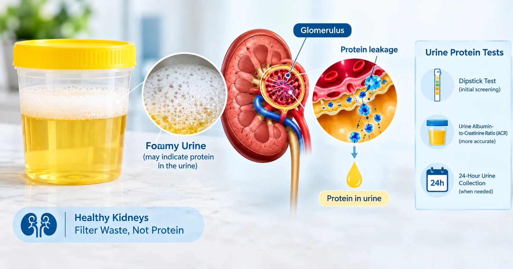
Reviewed by Dr. Jyoti Bansode, MBBS, MD Medicine, DM Nephrology (10+ years experience). Special interest: glomerular diseases. Seeing bubbles or a persistent foam on the surface of your urine can be unsettling. While occasional bubbles are normal, a consistently foamy stream may be a red flag for protein leakage—a hallmark of kidney dysfunction. What Is Foamy Urine and Why It Matters? Foamy urine is defined as a dense layer of small bubbles that remains on the surface of the toilet water for several seconds after voiding. Persistent foam often reflects excess protein (proteinuria) or, less commonly, blood (hematuria) in the urine, both of which can point to underlying kidney disease. According to a 2019 clinical review, persistent foam is considered a warning sign when it appears repeatedly and does not disappear quickly clinical study on foamy urine. Common Causes and Risk Factors Glomerular diseases such as minimal change disease, focal segmental glomerulosclerosis, or membranous nephropathy. Diabetes mellitus – high blood sugar damages the filtering units of the kidneys diabetes and kidney damage. Hypertension – long‑term high blood pressure strains glomeruli. Infections or inflammation of the urinary tract. Dehydration or rapid urine flow that traps air. Use of certain cleaning agents in the toilet that create artificial bubbles. Symptoms and Warning Signs Persistent foamy or bubbly urine (more than a few times a week). Swelling in the ankles, feet, or around the eyes (edema). Fatigue, especially after meals. Blood in the urine (hematuria) – visible pink or red tint. Decreased urine output or a feeling of incomplete emptying. High blood pressure that is difficult to control. How Doctors Evaluate Foamy Urine Urine Protein Tests The first step is a simple dip‑stick test that detects protein. For a more accurate assessment, clinicians order a urine albumin‑to‑creatinine ratio (ACR) or a 24‑hour urine protein collection. An ACR >30 mg/g is considered abnormal and may signal early kidney damage. Blood Tests and Imaging Blood work includes serum creatinine and estimated glomerular filtration rate (eGFR) to gauge overall kidney function. Imaging such as renal ultrasound can rule out structural causes like obstruction or cysts. Interpreting Test Results Normal vs. Pathologic Findings Occasional mild proteinuria (300 mg/g, especially when accompanied by hematuria, warrants further investigation for glomerular disease. When Findings Indicate Kidney Disease If the urine ACR is in the “moderate” (30‑300 mg/g) or “high” (>300 mg/g) range, and eGFR is below 60 mL/min/1.73 m², the pattern aligns with chronic kidney disease (CKD). In such cases, early intervention—blood pressure control, glycemic management, and possibly ACE inhibitors or ARBs—can slow progression. When to Seek Specialist Care Primary‑care providers often manage early kidney changes, but you should see a nephrologist when any of the following occur: Proteinuria persists on repeat testing despite lifestyle changes. eGFR falls below 60 mL/min/1.73 m². Visible blood in the urine or unexplained swelling. Blood pressure remains uncontrolled (>140/90 mmHg) while on three antihypertensives. You have a known risk factor such as diabetes, hypertension, or a family history of kidney disease. For a deeper dive into kidney‑function testing, read our guide on the <a href=”eGFR test and learn why high creatinine levels matter in <a href=”high creatinine causes. If you’re concerned about protein and blood in your urine, the detailed explanation on <a href=”foamy urine kidney disease provides a solid foundation. Practical Steps You Can Take Today Stay well‑hydrated; aim for at least 2 L of water daily. Limit high‑protein diets if you already have proteinuria; discuss with a dietitian. Control blood pressure and blood sugar aggressively. Schedule a repeat urine ACR in 2‑4 weeks to confirm persistence. Keep a symptom diary—note foam frequency, any swelling, and blood pressure readings. For additional reading on how foamy urine relates to broader kidney health, see the overview from HealthGrades and the patient‑focused guide from the Cleveland Clinic. Frequently Asked Questions Is occasional foamy urine normal? Yes, a single layer of bubbles that disappears quickly after a forceful stream is usually harmless and caused by air mixing with urine. Proteinuria above 30 mg/g on a urine ACR, especially if it persists on repeat testing, should be evaluated for kidney disease. Can dehydration cause foamy urine? Dehydration concentrates urine, which can increase foam, but the foam typically clears quickly; persistent foam still warrants testing. How quickly should I see a doctor if I notice blood in my urine? Any visible blood (hematuria) should prompt a medical visit within 24‑48 hours to rule out infection, stones, or kidney disease. Do over‑the‑counter supplements cause foamy urine? Some supplements, especially high‑dose vitamin C or protein powders, can temporarily increase urine protein, but persistent foam still requires evaluation. Is a low‑salt diet helpful for foamy urine? Reducing sodium can lower blood pressure and reduce kidney strain, which may help lessen protein leakage over time. Is a low‑salt diet helpful for foamy urine? Reducing sodium can lower blood pressure and reduce kidney strain, which may help lessen protein leakage over time. Conclusion If foamy urine appears repeatedly, it’s worth investigating with a urine protein test and basic kidney labs. Early detection can prevent irreversible damage, so schedule a check‑up promptly.