+91 8691888178 / 8850999397 dr.jyotibansode.01@gmail.com Monday to Saturday : 05:00PM TO 06:00PM

Blog

Diabetic Kidney Disease with Normal Creatinine – What You Need to Know

Yes, diabetic kidney disease can be present even when serum creatinine is within the normal laboratory range. Creatinine alone may miss early kidney damage because albumin leakage in urine often appears before a significant fall in the kidneys’ filtering ability. Why Creatinine Can Remain Normal in Early Diabetic Kidney Disease Serum creatinine reflects the overall filtration capacity of the kidneys, but it does not detect subtle damage to the glomerular filter. In the early stages of diabetic nephropathy, the glomerular basement membrane becomes leaky, allowing albumin to pass into the urine while the total filtration rate (and thus creatinine) stays within normal limits. Therefore, a patient can have: Normal creatinine Relatively preserved eGFR Elevated urine albumin (high urine ACR) and still have evidence of kidney damage. How Diabetes Damages the Glomeruli Prolonged high blood glucose damages the tiny blood vessels that line the glomeruli – the kidney’s filtering units. The excess glucose triggers inflammation and thickening of the basement membrane, making it porous. The first sign is usually a small amount of albumin spilling into the urine (microalbuminuria). Over time, continued injury can reduce the number of functional nephrons, leading to a drop in eGFR and, eventually, chronic kidney disease. Creatinine vs eGFR vs Urine ACR Each test answers a different question about kidney health. Understanding their roles helps catch disease early. Test What It Measures Typical Use Creatinine Blood waste product from muscle metabolism Basic screen; high levels suggest reduced filtration eGFR Estimated glomerular filtration rate (calculates filtration based on creatinine, age, sex, race) Quantifies overall kidney function Urine ACR Albumin‑to‑creatinine ratio in a spot urine sample Detects early leak of protein (albumin) from glomeruli Because they assess different aspects, clinicians interpret them together rather than in isolation. Can eGFR Be Normal in Early Diabetic Kidney Disease? Yes. In the initial phases of diabetic nephropathy, the eGFR often remains >90 mL/min/1.73 m² while urine ACR is already elevated. A normal eGFR does not guarantee that the kidneys are healthy; persistent albuminuria signals early injury even when filtration appears preserved. Current Screening Guidance for Diabetes Guidelines from the American Diabetes Association and KDIGO recommend: People with type 2 diabetes: urine ACR and eGFR testing at least once a year from the time of diagnosis. People with type 1 diabetes: begin annual kidney screening after about five years of diabetes duration, unless other risk factors call for earlier testing. Regular monitoring helps detect changes before symptoms appear. Early Warning Signs (Often Absent) Early diabetic kidney disease usually has no noticeable symptoms. When it progresses, patients may notice: Foamy or frothy urine (protein) Swelling of the feet or ankles Increasing blood pressure Fatigue or reduced energy Changes in urination frequency Nausea or loss of appetite Can a High Urine ACR Be Temporary? Transient factors can raise urine albumin, including intense exercise, fever, urinary‑tract infection, marked hyperglycaemia, menstruation, heart failure, or a sudden spike in blood pressure. One abnormal ACR result does not automatically confirm chronic kidney disease; repeat testing after a few weeks is recommended to verify persistence. What Test Results May Suggest Early Kidney Damage? Look for patterns such as: Normal serum creatinine + elevated urine ACR. Preserved eGFR + elevated urine ACR. Gradual decline in eGFR over months or years. Increasing albumin levels on consecutive ACR tests. These findings indicate that the kidneys are under stress even if the filtration rate seems normal. What Should You Do If Creatinine Is Normal but You Have Diabetes? Discuss the following with your clinician: Request a urine ACR test. Ask for an eGFR calculation based on your latest labs. Review previous creatinine results to spot trends. Review prior urine ACR results, if available. Check your blood pressure readings. Know your most recent HbA1c level. Ask whether repeat urine testing is needed to confirm any abnormality. Inquire if your kidney‑function numbers are changing over time. When Should You See a Nephrologist? Referral is appropriate when any of the following occur: Persistently elevated urine ACR (especially >300 mg/g). Rapid increase in albuminuria over a short period. Steady decline in eGFR (e.g., drop >5 mL/min/1.73 m² per year). Persistent protein or blood in urine protein and blood in urine. Difficult‑to‑control hypertension despite medication. Unexplained swelling (edema). Sudden or unexplained changes in kidney‑function test results. Uncertainty about the cause of abnormal kidney tests acute and chronic kidney diseases. Referral does not mean you have end‑stage disease; it ensures specialized care to slow progression. Why Early Detection Matters Interventions such as tighter glucose control, blood‑pressure management (often with ACE inhibitors or ARBs), lifestyle changes, and regular monitoring can significantly slow the progression of diabetic kidney disease. Medical Review: Dr. Jyoti BansodeMBBS, MD Medicine, DM NephrologyMore than 10 years of nephrology experience For a deeper look at how diabetes affects kidney health, see our diabetes and kidney diseases page. If you notice persistent protein or blood in your urine, read our protein and blood in urine guide. For broader information on chronic kidney conditions, explore acute and chronic kidney diseases. Key sources:  American Heart Association overview of kidney disease in diabetes, bidirectional link study between diabetes and kidney disease, KDIGO‑style review of diabetic kidney disease. Frequently Asked Questions Can diabetic kidney disease happen with normal creatinine? Yes. Early kidney damage can cause albumin to leak into urine while serum creatinine remains within the normal range. Can kidney damage occur with a normal eGFR? Yes. Albuminuria can be present even when eGFR is >90 mL/min/1.73 m², indicating early glomerular injury. Which test detects diabetic kidney disease earliest? Urine ACR (albumin‑to‑creatinine ratio) is the most sensitive early marker, detecting microalbuminuria before creatinine or eGFR change. Is urine ACR more useful than creatinine for early kidney damage? For early detection, yes. Urine ACR identifies tiny amounts of albumin that appear before serum creatinine rises. Can albumin in urine be temporary? Transient factors like exercise, infection, fever, or high blood pressure can raise urine albumin; repeat testing confirms persistence. When should a person with diabetes see a nephrologist? See a nephrologist if urine ACR stays high, eGFR declines, protein/blood persists in urine, hypertension is uncontrolled,

Protein in Urine Causes: What’s Normal, What’s Worrisome?

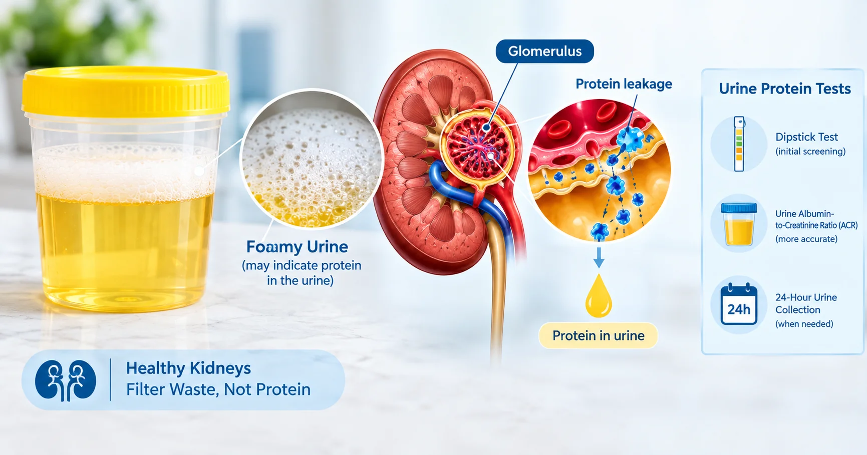
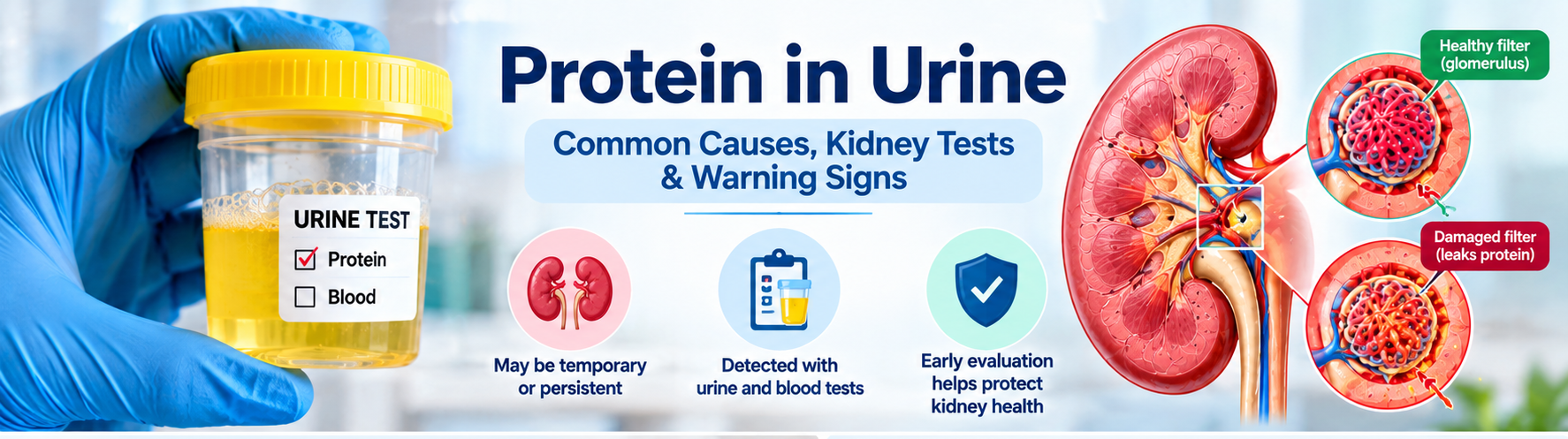
Short answer : Protein in urine can be a fleeting finding after fever, intense exercise, or dehydration, but persistent proteinuria often signals damage to the kidney’s filtering units (glomeruli). Ongoing leakage may require further testing and specialist care. Proteinuria—often discovered on a routine urine test—doesn’t always mean serious disease. A single abnormal result may be temporary, yet repeated or high‑level protein loss usually points to kidney‑filter damage that needs evaluation. What Is Proteinuria? Healthy kidneys keep large proteins such as albumin inside the bloodstream while allowing waste and excess fluid to pass into urine. The glomeruli act as tiny sieves; when they are inflamed, scarred, or otherwise damaged, proteins slip through and appear in the urine. How the kidneys normally work  – Blood enters the glomerulus at high pressure.  – Small molecules (water, electrolytes, waste) filter out. – Large proteins are retained and re‑absorbed in the tubules. When the glomerular barrier is compromised, albumin and other proteins are lost, leading to a measurable proteinuria. Common Causes of Protein in Urine (Persistent) These conditions often produce ongoing protein leakage and merit a thorough work‑up:  – Diabetes mellitus – high blood sugar damages glomerular capillaries.  – Hypertension (high blood pressure) – pressure overload injures the filtration barrier.  – Glomerular diseases – e.g., IgA nephropathy, membranous nephropathy, focal segmental glomerulosclerosis.  – Chronic infections – hepatitis C, HIV, or recurrent urinary‑tract infections.  – Cardiovascular conditions – heart failure can raise kidney venous pressure.  – Medications – non‑steroidal anti‑inflammatory drugs (NSAIDs), certain antibiotics, and some chemotherapy agents.  – Other kidney disorders – polycystic kidney disease, obstructive uropathy. For an in‑depth look at these factors, see Causes of Protein in Urine: Understanding the Key Factors. Temporary Causes of Protein in Urine Not every positive protein test signals chronic disease. Transient proteinuria often resolves once the trigger disappears: Fever or acute illness – inflammation raises glomerular permeability. Strenuous exercise – muscle breakdown and increased blood flow can spill protein. Dehydration – concentrated urine may falsely appear protein‑rich. Cold exposure – vasoconstriction can temporarily affect filtration. Recent urinary‑tract infection – inflammation of the bladder or urethra. Because a single abnormal result may be fleeting, clinicians often repeat a morning urine sample before ordering extensive work‑up. Symptoms and Warning Signs Early proteinuria is frequently silent, but watch for these clues:  – Foamy, frothy, or bubbly urine.  – Swelling (edema) of ankles, feet, or around the eyes.  – Unexplained fatigue or loss of appetite.  – Changes in urine volume or frequency.  – Elevated blood pressure. Most people with mild proteinuria feel fine; the presence of symptoms often indicates a higher level of protein loss or concurrent kidney dysfunction. Does foamy urine always mean protein in urine? No. Foam can result from rapid urine flow, concentrated urine, or even certain detergents in the toilet bowl. Only a laboratory test (dipstick or ACR) can confirm proteinuria. Protein in Urine vs. Blood in Urine Proteinuria (protein) and hematuria (blood) are distinct findings. Protein indicates a leak of albumin or larger proteins, while blood signals red‑cell leakage, often from a different part of the urinary tract. When both appear together, clinicians consider glomerular disease or severe inflammation. Read more about the overlap in our protein and blood in urine article. How Proteinuria Is Measured The most common tests include: – Urine dipstick – quick screening, semi‑quantitative.  – Urine albumin‑to‑creatinine ratio (UACR) – expresses albumin in mg per gram of creatinine; preferred for diabetes monitoring.  – Urine protein‑to‑creatinine ratio (PCR) – measures total protein.  – Comprehensive urinalysis – evaluates sediment, cells, and casts.  – Serum creatinine & eGFR – assess overall kidney filtration function. Interpretation of UACR:Range (mg/g)Interpretation<30Normal or low‑grade30–300Micro‑albuminuria (early kidney damage)>300Macro‑albuminuria (significant kidney disease) Urine ACR (UACR) Result Interpretation UACR Result Interpretation Below 30 mg/g Normal to mildly increased 30–300 mg/g Moderately increased albuminuria Above 300 mg/g Severely increased albuminuria Results should be repeated, especially if the first sample was a random collection, and interpreted alongside creatinine, eGFR, and clinical context. Can You Have Protein in Urine with Normal Creatinine? Yes. Protein leakage can precede a rise in serum creatinine because creatinine reflects overall filtration, not the selective loss of albumin. Early glomerular injury may show a high UACR while eGFR remains >90 mL/min/1.73 m². Glomerular Disease: When to Be Concerned Persistent, significant proteinuria—especially when accompanied by any of the following—should raise suspicion for a primary glomerular disorder:  – Protein levels >300 mg/g (macro‑albuminuria).  – Concurrent hematuria (blood in urine).  – Unexplained edema or high blood pressure.  – Declining eGFR over months.  – Abnormal urine sediment (casts, dysmorphic RBCs). Dr. Jyoti Bansode has a special clinical interest in glomerular diseases; patients with these red flags are often referred for nephrology evaluation. When Might a Kidney Biopsy Be Needed? Most proteinuria cases are managed without invasive procedures. A biopsy is considered when:  – The underlying cause remains unclear after standard labs and imaging.  – Protein loss exceeds 3.5 g/day (nephrotic‑range) with rapid eGFR decline.  – There is suspicion of a specific glomerular disease that guides therapy (e.g., lupus nephritis, IgA nephropathy).  – Patients have refractory proteinuria despite optimal blood‑pressure and diabetes control. Read more about the procedure in our kidney biopsy overview. Patient Checklist After an Abnormal Urine Protein Test Ask which test was performed (dipstick, UACR, PCR). Confirm the exact protein value and its units. Request your serum creatinine and eGFR results. Discuss any recent fever, intense exercise, dehydration, or infection that could be temporary. Inquire whether a repeat morning urine sample is recommended. Review blood‑pressure control and diabetes management. Ask if a referral to a nephrologist is appropriate. When to See a Nephrologist Consider specialist care if you have any of the following:  – Persistent proteinuria on two separate tests (≥30 mg/g).  – Rising UACR or PCR over weeks to months.  – Proteinuria accompanied by hematuria.  – Declining eGFR (<60 mL/min/1.73 m²) or rising serum creatinine.  – Significant edema or uncontrolled hypertension.  – Suspected glomerular disease based on urine sediment.  – Unclear cause after standard work‑up. Schedule a consultation through our appointment and consultation page. Medical ReviewDr. Jyoti

Diabetic Kidney Disease Symptoms: Early Signs & Tests

Diabetic kidney disease (DKD), also called diabetic nephropathy, is a common complication of diabetes. Current American Diabetes Association guidance estimates that chronic kidney disease affects about 20–40% of people with diabetes. The condition may develop without noticeable symptoms, so regular urine and blood testing is important for detecting kidney damage early.  Medical Review: Dr. Jyoti Bansode, MBBS, MD Medicine, DM Nephrology — more than 10 years of nephrology experience. Quick Answer: What Are the Early Symptoms of Diabetic Kidney Disease? Early diabetic kidney disease often causes no obvious symptoms. The first signs may appear on kidney tests, such as increased albumin in urine or changes in eGFR. As kidney disease progresses, possible symptoms may include:  – Foamy or unusually bubbly urine  – Swelling in the feet, ankles or around the eyes  – Fatigue or weakness  – Changes in urination  – Nausea or reduced appetite  – Itching or muscle cramps  – Increasing or difficult-to-control blood pressure Because early diabetic kidney disease is often silent, blood and urine tests are more reliable than waiting for symptoms to appear.  What Is Diabetic Kidney Disease? Diabetic kidney disease develops when diabetes damages the small blood vessels and filtering structures inside the kidneys. The kidneys contain microscopic filtering units called glomeruli. Over time, persistently high blood glucose can damage these filters, allowing albumin to leak into the urine and gradually reducing the kidneys’ ability to remove waste from the blood. High blood pressure can further increase stress on the kidneys and accelerate kidney damage. For a broader explanation, read about diabetes and kidney diseases. Why Can Diabetic Kidney Disease Develop Without Symptoms? Kidneys have significant reserve capacity. Early kidney damage may therefore develop while a person still feels completely well. NIDDK notes that most people with diabetic kidney disease do not have symptoms in the early stages. Kidney problems are usually detected through blood and urine tests.  A patient can have: No pain + no swelling + normal-looking urine and still have albuminuria or another early kidney abnormality. Who Is at Higher Risk? Not everyone with diabetes develops diabetic kidney disease. Risk may be greater in people who have: Diabetes for many years Persistently high blood glucose High blood pressure Cardiovascular disease Smoking habits Excess body weight Family history of kidney failure Existing kidney abnormalities NIDDK identifies poor blood-glucose control and high blood pressure as especially important risk factors.  Type 1 vs Type 2 Diabetes: When Should Kidney Testing Begin? Type 1 Diabetes Current ADA 2026 recommendations advise checking urine ACR and eGFR at least annually once a person has had type 1 diabetes for five years or more.  Type 2 Diabetes People with type 2 diabetes should generally have urine ACR and eGFR checked at least once a year from diagnosis, regardless of treatment. Diabetes Journals This difference matters because type 2 diabetes may have been present for years before it is diagnosed. What Are the Earliest Signs of Kidney Damage in Diabetes? The earliest signs are often found on laboratory tests. 1. Albumin in the Urine Albumin is a protein that normally remains in the bloodstream. When the kidney filters are damaged, albumin can begin leaking into urine. This is called albuminuria. The standard test used to measure this is the urine albumin-to-creatinine ratio (UACR). For more detail, see the Urine ACR Test Guide. 2. Changes in eGFR The estimated glomerular filtration rate (eGFR) estimates how well the kidneys are filtering blood. A declining eGFR can indicate reduced kidney function. 3. Changes in Serum Creatinine Creatinine is a waste product normally filtered from the blood by the kidneys. As filtration worsens, serum creatinine may increase. However, creatinine alone is not enough to assess early diabetic kidney disease. 4. Increasing Blood Pressure High blood pressure can damage kidney blood vessels, while kidney disease can also make blood pressure more difficult to control. Patients with persistent blood-pressure problems can read more about resistant hypertension. The Two Most Important Tests for Diabetic Kidney Disease The two key kidney markers are: Urine ACR (UACR) eGFR NIDDK specifically identifies urine albumin and eGFR as the two major markers used to detect and monitor chronic kidney disease.  Urine Albumin-to-Creatinine Ratio (UACR) UACR measures albumin in urine relative to creatinine. A random spot urine sample is usually sufficient, meaning a 24-hour urine collection is generally unnecessary for routine screening.  Typical categories include: UACR Result Interpretation UACR Result Interpretation Below 30 mg/g Normal to mildly increased 30–300 mg/g Moderately increased albuminuria Above 300 mg/g Severely increased albuminuria A UACR above 30 mg/g is considered abnormal, but the result should be interpreted with other kidney tests and, when appropriate, repeat measurements.  eGFR eGFR estimates kidney filtration. In general: 60 or above: may be within an acceptable range depending on the clinical situation Below 60: may indicate kidney disease if the reduction persists 15 or below: represents severely reduced kidney function and may indicate kidney failure The result should always be interpreted alongside urine albumin and the patient’s medical history. Can You Have Diabetic Kidney Disease with a Normal eGFR? Yes. A person may have: Normal or relatively preserved eGFR + elevated urine ACR and still have evidence of kidney damage. Albuminuria can appear before significant changes in eGFR. This is why checking only creatinine or eGFR may miss early kidney damage.  How to Understand UACR and eGFR Together How to Understand UACR and eGFR Together UACR eGFR What It May Suggest Normal Normal Current kidney markers may be reassuring. High Normal Possible early kidney damage. Normal Low Reduced kidney filtration requiring evaluation. High Low Kidney damage with reduced filtration may be present. This table is a general guide only. Diagnosis depends on repeat testing, trends over time and the patient’s overall clinical condition. Learn more about combined kidney testing in Kidney Tests Explained. Can a High Urine ACR Be Temporary? Yes. A single elevated urine ACR does not always mean permanent kidney damage. Factors that can temporarily raise UACR include: – Vigorous exercise within the previous 24 hours  – Infection  –

Urine ACR Test Explained: Why Albumin in Urine Matters

When your doctor orders a urine albumin‑creatinine ratio (ACR) test, the goal is to catch early signs of kidney stress before symptoms appear. This guide breaks down the science in plain language, explains what the numbers mean, and shows you how to prepare for accurate results. Medical Review: Dr. Jyoti Bansode, MBBS, MD Medicine, DM Nephrology – >10 years of nephrology experience. What the Urine ACR Test Measures The ACR test quantifies two substances in a single, random‑spot urine sample: albumin (a protein that belongs in the bloodstream) and creatinine (a waste product filtered by the kidneys). The laboratory divides the albumin concentration (mg) by the creatinine concentration (g) to produce a ratio expressed as mg/g. Because creatinine corrects for urine concentration, the ACR is more reliable than a simple dip‑stick protein test. For a detailed explanation, see Dr. Jyoti Bansode’s article. Albumin vs. Creatinine Albumin should stay in the blood; the kidneys act as a sieve that prevents it from leaking into urine. Creatinine, on the other hand, is a by‑product of muscle metabolism that the kidneys normally excrete. By comparing the two, the ACR tells you whether the filtration barrier is compromised. Why Albumin in Urine Matters Even tiny amounts of albumin (called microalbuminuria) signal that the glomeruli—the tiny filtering units—are under stress. Persistent albuminuria is linked to a higher risk of chronic kidney disease (CKD), cardiovascular events, and progression to end‑stage renal disease. The National Kidney Foundation notes that albuminuria is a red flag for both kidney and heart health NKF uACR overview. Understanding ACR Result Ranges Normal: < 30 mg/g – no significant albumin loss. Microalbuminuria: 30–300 mg/g – early kidney damage; warrants repeat testing and risk‑factor control. Macroalbuminuria: >300 mg/g – overt proteinuria; indicates more advanced disease. These cut‑offs are widely accepted in clinical practice and help guide treatment decisions NKF uACR overview. Common Causes of a High ACR Diabetes mellitus – high blood sugar damages glomerular capillaries. Hypertension – elevated pressure strains the filtration barrier. Glomerulonephritis or other inflammatory kidney diseases. Infections (e.g., urinary tract infection, fever). Medications such as non‑steroidal anti‑inflammatory drugs (NSAIDs) or certain antibiotics. For a deeper look at how diabetes triggers albuminuria, read our Understanding Microalbuminuria in Diabetes. Temporary Factors That Can Skew Results Not every elevated ACR means chronic disease. Transient conditions can raise albumin temporarily: Intense exercise or heavy lifting (muscle breakdown releases creatinine). Fever or acute illness. Dehydration – concentrates urine, raising the ratio. High‑protein meals within the previous 24 hours. Menstrual blood contamination. If any of these apply, your clinician may repeat the test after a few days. Interpreting ACR with eGFR and Creatinine ACR is most powerful when viewed alongside estimated glomerular filtration rate (eGFR) and serum creatinine. A normal eGFR with a high ACR suggests early glomerular injury, while a low eGFR with a modest ACR may reflect advanced CKD. Our Kidney Tests Explained: Creatinine, eGFR, Urine ACR & Electrolytes article walks through the combined interpretation. Laboratories such as Labcorp calculate the ratio automatically and flag results that exceed risk thresholds Labcorp ACR test details. Diabetes, Hypertension, and Cardiovascular Risk Albuminuria is not just a kidney issue; it predicts heart attacks, strokes, and peripheral artery disease. People with diabetes and high blood pressure are up to three times more likely to develop albuminuria, and each 30 mg/g increase in ACR raises cardiovascular risk by roughly 10 % according to the National Kidney Foundation NKF uACR risk page. How Often Should You Repeat the Test? Guidelines recommend: Annual screening for anyone with diabetes or hypertension. Every 1–2 years for individuals with other CKD risk factors (family history, age > 60). Repeat within 3–6 months if the first result is abnormal, after addressing any temporary factors. When to See a Nephrologist Schedule a specialist appointment if you meet any of the following: ACR > 300 mg/g on two separate occasions. eGFR < 60 mL/min/1.73 m² combined with any albuminuria. Rapidly rising ACR (increase > 30 % within 3 months). Persistent proteinuria despite optimal blood‑pressure and glucose control. Our guide on specialist referral can help you prepare for the visit: When Should You See a Nephrologist in Navi Mumbai?. Practical Patient Checklist Before Your ACR Test Collect a mid‑stream, clean‑catch urine sample in the morning if possible. Avoid vigorous exercise for 24 hours prior. Stay well‑hydrated, but do not over‑drink immediately before the test. Notify the lab if you have a fever, infection, or recent heavy protein intake. Bring a list of current medications (especially NSAIDs, ACE inhibitors, or ARBs). Following these steps improves accuracy and reduces the need for repeat testing. Frequently Asked Questions What does a normal urine ACR result look like? A normal ACR is less than 30 mg of albumin per gram of creatinine, indicating that the kidneys are effectively retaining protein. Can a single high ACR result be ignored? No. A single elevated ACR should be repeated after 3–6 months, especially if you had recent fever, intense exercise, or dehydration that could temporarily raise the ratio. Why is the ACR preferred over a simple urine protein dip‑stick? The ACR corrects for urine concentration by using creatinine as a reference, making it more reliable than a dip‑stick, which can be falsely low in dilute urine or high in concentrated samples. How does diabetes affect the urine ACR? High blood glucose damages the glomerular filter, allowing albumin to leak into urine. Even early diabetes can cause microalbuminuria (30–300 mg/g), which signals the need for tighter glucose and blood‑pressure control. When should I see a nephrologist after an abnormal ACR? If your ACR exceeds 300 mg/g, if eGFR falls below 60 mL/min/1.73 m², or if albuminuria rises rapidly despite treatment, a referral to a nephrologist is recommended. Conclusion Understanding your urine ACR result empowers you to act early, protect kidney function, and lower cardiovascular risk. Talk to your healthcare provider about the next steps, whether that means lifestyle changes, medication adjustments, or a referral to a nephrologist.

Urine ACR Test Explained: Why Albumin in Urine Matters

The urine albumin‑creatinine ratio (urine ACR test) is a quick, inexpensive way to spot early kidney damage before symptoms appear. By comparing the amount of albumin (a protein) to creatinine (a waste product) in a single urine sample, doctors can detect albuminuria, monitor disease progression, and guide treatment for conditions like diabetes and hypertension. What Is a Urine ACR Test? A urine ACR test measures the concentration of albumin and creatinine in a spot urine sample and then calculates the ratio, usually expressed in milligrams of albumin per gram of creatinine (mg/g). This ratio corrects for variations in urine concentration, making it more reliable than a simple urine protein test. According to the National Kidney Foundation, the uACR is the preferred screening tool for detecting albuminuria in people at risk for chronic kidney disease (CKD). Why Is the Urine ACR Test Done? Kidneys filter waste while keeping essential proteins like albumin in the bloodstream. When the glomeruli become damaged, albumin leaks into the urine, a condition called albuminuria or proteinuria. Detecting this leak early can: Identify early-stage CKD before eGFR declines. Stratify cardiovascular risk, since albuminuria is an independent predictor of heart disease. Guide medication choices for diabetes, hypertension, and other kidney‑affecting conditions. For people with diabetes, the Kidney Fund notes that routine uACR screening is essential because diabetic kidney disease often begins with microalbuminuria. How Is a Urine ACR Test Done? The test requires a clean‑catch midstream urine sample, which can be collected at home or in a clinic. No fasting is needed, but the sample should be taken when the bladder is neither empty nor overly full. The laboratory measures albumin (mg) and creatinine (g) and reports the ratio. Some labs also provide a separate urine albumin test and a urine creatinine test, but the combined ratio is what clinicians use for interpretation. Understanding Urine ACR Results Results are categorized by the National Kidney Foundation as follows: Understanding urine ACR result with kidney illustration and normal, moderately increased, and severely increased UACR ranges UACR (mg/g) Albuminuria Category What It Means < 30 mg/g Normal to Mildly Increased Usually considered within the normal range. 30–299 mg/g Moderately Increased May indicate early kidney damage or albuminuria. ≥ 300 mg/g Severely Increased May indicate more significant kidney damage and needs medical evaluation. A result under 30 mg/g is considered normal, while values between 30 and 299 mg/g indicate microalbuminuria—a warning sign that kidney damage may be beginning. Values of 300 mg/g or higher reflect macroalbuminuria, which usually means more advanced kidney disease. It’s important to remember that a single abnormal result does not automatically diagnose CKD. The NIDDK recommends repeating the urine ACR test one to two more times over a 3‑month period to confirm persistent albuminuria before labeling someone with chronic kidney disease. UACR vs. Other Urine Protein Tests Traditional urine protein tests measure total protein, which can be influenced by hydration status and muscle mass. The urine ACR test, by normalizing albumin to creatinine, provides a more stable estimate of kidney filtration function. This is why clinicians often prefer the uACR over a generic urine protein test when monitoring diabetic patients or those with hypertension. Key Situations When a Urine ACR Test Is Recommended How is a urine ACR test done showing urine collection, albumin measurement, creatinine measurement, and UACR calculation Annual screening for adults with diabetes or hypertension. Evaluation of unexplained swelling, foamy urine, or decreased urine output. Baseline assessment before starting potentially nephrotoxic medications (e.g., NSAIDs, certain antibiotics). Follow‑up after a previous abnormal ACR to assess treatment response. For a deeper dive into related kidney tests, see our Kidney Tests Explained: Creatinine, eGFR, Urine ACR & Electrolytes. Interpreting High Urine ACR Results When the urine ACR is above 30 mg/g, clinicians consider several possible causes: Early diabetic nephropathy or other forms of CKD. Acute conditions such as urinary tract infection, fever, or intense exercise. Medications that affect kidney function. Addressing reversible factors—like treating an infection or adjusting medication—can sometimes bring the ACR back into the normal range. Lifestyle changes (tight blood‑pressure control, low‑protein diet, smoking cessation) also help lower albuminuria over time. Link Between UACR and eGFR eGFR estimates how well the kidneys filter blood, while uACR detects leakage of protein. Both are complementary: a normal eGFR with a high uACR signals early kidney damage, whereas a low eGFR with a normal uACR may indicate non‑proteinuric CKD. Combining these metrics improves risk stratification, as highlighted by the Labcorp ACR test description. Practical Tips for Accurate Testing To ensure reliable results, follow these guidelines: Collect the sample first thing in the morning, if possible. Avoid heavy exercise, high‑protein meals, and NSAIDs for 24 hours before the test. Store the sample in a cool place and deliver it to the lab within 24 hours. Inform your provider about any recent infections or medications. For more on how to prepare for lab tests, read our Protein in Urine Child: When to Seek Kidney Care. When to Repeat the Test If your first urine ACR result is abnormal, most clinicians will repeat the test after 2–4 weeks to confirm persistence. For patients with known CKD, the test may be ordered every 3–6 months, depending on disease stability and treatment changes. Diabetic patients are often advised to have a urine ACR at least once a year, as recommended by the American Diabetes Association. Can High Urine ACR Be Reversed? Yes, in many cases. Tight glycemic control, blood‑pressure management (especially with ACE inhibitors or ARBs), and lifestyle modifications have been shown to reduce albuminuria. Studies cited by the Siemens Healthineers indicate that up to 30 % of patients can achieve a return to the normal range with appropriate therapy. Ultimately, early detection through regular urine ACR screening empowers patients and providers to intervene before irreversible kidney damage occurs. Frequently Asked Questions What is a urine ACR test? A urine ACR test measures the albumin‑to‑creatinine ratio in a spot urine sample, providing a snapshot of how much albumin is leaking into the urine relative to creatinine. What does a high

Understanding Microalbuminuria in Diabetes: What an Abnormal Urine Test Means

When you receive a lab report that says your urine albumin is elevated, it can be unsettling. For people with diabetes, an abnormal urine test often points to microalbuminuria – a subtle but important sign of early kidney stress. This article explains what microalbuminuria in diabetes means, how the test is performed, how to interpret the numbers, and what steps you can take to protect your kidneys. What Is Microalbuminuria in Diabetes? Microalbuminuria refers to a moderate increase in the amount of albumin – a small protein – that leaks into the urine. In healthy kidneys, less than 30 mg of albumin is excreted per gram of creatinine. When the level rises to 30–300 mg/g, the condition is called microalbuminuria; values above 300 mg/g are termed macroalbuminuria or overt proteinuria. According to early diabetes research, this rise is often invisible on a standard dipstick and requires a specific urine microalbumin test. Why It Matters for People With Diabetes Elevated urine albumin is the first measurable sign that the tiny filtering units (glomeruli) in the kidneys are being damaged by high blood sugar. It also signals a higher risk of cardiovascular events. Detecting microalbuminuria early gives you a window to intervene before diabetic nephropathy becomes irreversible. How the Urine Microalbumin Test Works The most common screening method is a spot urine sample that measures both albumin and creatinine, then calculates the urine albumin‑to‑creatinine ratio (UACR). This ratio corrects for variations in urine concentration, making it reliable even when you haven’t timed the collection. Sample Collection Options Random spot sample: The easiest method; you provide a single urine specimen at any time of day. First‑morning sample: Slightly more concentrated, reducing variability. 24‑hour collection: Considered the gold standard but is cumbersome for most patients. Most guidelines recommend an annual spot‑sample screening for adults with diabetes aged 12–70 years clinical recommendation. Understanding the UACR The UACR is reported in milligrams of albumin per gram of creatinine (mg/g). The interpretation is straightforward: Normal: < 30 mg/g Microalbuminuria: 30–300 mg/g (equivalent to 30–300 mg albumin in a 24‑hour collection) Macroalbuminuria: > 300 mg/g These cut‑offs match the definitions used in the Ubie Doctor’s note and are echoed in the MLabs reference. Interpreting Test Results: More Than Just Numbers Understanding UACR result with urine sample, laboratory analysis, and albuminuria interpretation ranges When you see a result labeled “high urine albumin,” consider the following factors before concluding permanent kidney damage: Temporary physiological changes: Fever, vigorous exercise, or a high‑protein meal can transiently raise albumin excretion. Medications: Certain drugs (e.g., NSAIDs) may affect kidney filtration. Laboratory variability: Different labs use slightly different units; always confirm the reference range. If an abnormal value appears, clinicians typically repeat the test. Two out of three positive UACR results over a 3–6‑month period confirm true microalbuminuria clinical protocol. Common Causes of a One‑Time Elevated Albumin Level Before jumping to a diagnosis of diabetic nephropathy, rule out these reversible contributors: Acute urinary tract infection Uncontrolled hypertension Recent intense physical activity (e.g., marathon running) Dehydration or concentrated urine High‑protein diet within the past 24 hours Addressing these factors often normalizes the UACR on repeat testing. Confirming an Abnormal Result – The Screening Protocol Guidelines recommend the following workflow after a first abnormal microalbuminuria reading: Repeat the spot‑sample UACR in 1–3 months. If the second test is still elevated, obtain a third sample within the next 3 months. Two of the three results above 30 mg/g confirm persistent microalbuminuria. Once confirmed, a quantitative assessment of kidney function – including eGFR and serum creatinine – should be ordered. Our Kidney Tests Explained page walks through how eGFR and creatinine complement the urine albumin test. What Microalbuminuria Means for Your Kidneys Persistent microalbuminuria signals early diabetic kidney disease, often called diabetic nephropathy. Studies show that structural changes in the glomeruli can begin a decade before overt symptoms appear research on subclinical kidney changes. If left unchecked, microalbuminuria can progress to macroalbuminuria, a rapid decline in eGFR, and eventually end‑stage renal disease. Because albuminuria reflects systemic endothelial dysfunction, it also flags an increased risk for heart disease and stroke – a dual warning that warrants aggressive risk‑factor management. Protect your kidneys infographic showing blood sugar, blood pressure, diet, monitoring, and medical follow-up in diabetes Managing and Reducing Albuminuria Intervention at the microalbuminuria stage can halt or even reverse kidney damage. Key strategies include: Blood pressure control: Aim for <130/80 mm Hg using ACE inhibitors or ARBs, which specifically reduce intraglomerular pressure. Optimized glycemic control: Target an HbA1c below 7 % (or individualized per provider). Lifestyle modifications: Low‑sodium diet, regular moderate exercise, weight management, and smoking cessation. Lipid management: Statins lower cardiovascular risk, indirectly protecting kidney health. Regular monitoring—every 3–6 months for the first year after detection—helps gauge treatment effectiveness. If albuminuria persists despite optimal therapy, referral to a nephrologist is advised. When to See a Kidney Specialist Consider a referral if any of the following occur: Macroalbuminuria (> 300 mg/g) on two consecutive tests eGFR falls below 60 mL/min/1.73 m² Rapid rise in UACR despite ACE/ARB therapy Uncontrolled hypertension or diabetes despite standard measures Early involvement of a nephrologist can tailor medication regimens, discuss potential renal protective agents, and plan for future renal replacement therapy if needed. For more guidance on when specialist care is appropriate, see our dialysis criteria article. Integrating the Urine Test Into Your Diabetes Care Plan Think of the urine albumin test as a vital sign for your kidneys, just like blood pressure or blood sugar. Incorporate it into your routine diabetes check‑ups, discuss results openly with your endocrinologist, and track trends over time. By staying proactive, you can keep microalbuminuria from becoming a roadblock on your health journey. Frequently Asked Questions What is the normal range for urine albumin‑to‑creatinine ratio (UACR)? A normal UACR is less than 30 mg of albumin per gram of creatinine. Values between 30 and 300 mg/g indicate microalbuminuria, and above 300 mg/g suggest macroalbuminuria. How often should people with diabetes get a urine microalbumin test? Guidelines recommend an annual spot‑sample UACR for adults with diabetes aged 12–70. If microalbuminuria is detected, repeat testing every 3–6 months until the trend is clear. Can a

Kidney Tests Explained: Creatinine, eGFR, Urine ACR & Electrolytes

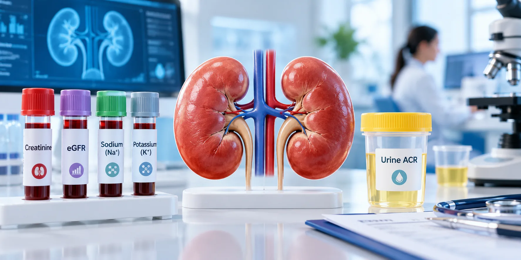
Kidney Tests Explained: Creatinine, eGFR, Urine ACR & ElectrolytesUnderstanding kidney function starts with the right lab tests. Creatinine, eGFR, urine albumin‑to‑creatinine ratio (uACR) and electrolyte panels are the four pillars doctors use to spot early disease, track progression, and guide treatment. This guide breaks down each test, normal values, and what the numbers mean for you. Medical Review – Dr. Jyoti BansodeMBBS, MD Medicine, DM NephrologyOver 10 years of experience in diagnosing and managing kidney disease. For personalized care, visit Dr. Bansode’s nephrology services. Why Kidney Function Testing Matters Kidneys filter waste, balance fluids, and regulate electrolytes. When they falter, waste builds up, blood pressure spikes, and vital minerals become imbalanced. Early detection through lab testing can prevent irreversible damage and reduce the need for dialysis or transplant. Common Symptoms That Prompt Testing Persistent fatigue or weakness Swelling in ankles, feet, or face Changes in urine color or frequency Unexplained high blood pressure Muscle cramps or tingling sensations If you notice any of these, ask your primary‑care physician for a kidney panel. Key Kidney Tests Overview Key Kidney Tests Overview Serum Creatinine – What It Shows Creatinine is a waste product from muscle metabolism. The kidneys clear it from the blood, so higher levels indicate reduced filtration. Normal ranges vary by sex and muscle mass, but typical limits are 0.6–1.2 mg/dL for women and 0.7–1.3 mg/dL for men Nephrology Specialists. Values above these thresholds often signal early kidney stress. Estimated Glomerular Filtration Rate (eGFR) eGFR translates the creatinine value into an estimate of how many milliliters of blood the kidneys filter each minute per 1.73 m² of body surface. An eGFR > 60 mL/min/1.73 m² is generally considered normal, 30–59 suggests moderate decline, and <30 indicates severe kidney disease MyNephrologist. The calculation also incorporates age, sex, and sometimes race, making it a more comprehensive indicator than creatinine alone. Urine Albumin‑to‑Creatinine Ratio (uACR) Albumin is a protein that should stay in the bloodstream. When the glomeruli are damaged, albumin leaks into urine. uACR compares the amount of albumin to creatinine in a single urine sample, eliminating the need for a 24‑hour collection. Normal uACR is 300 mg/g reflects macro‑albuminuria (significant damage) MyNephrologist. This test can detect kidney injury years before eGFR drops. Electrolytes and Blood Urea Nitrogen (BUN) Electrolyte panels measure sodium, potassium, chloride, bicarbonate, and calcium—minerals the kidneys tightly regulate. Abnormalities often accompany chronic kidney disease (CKD). BUN, another waste product, rises as kidney function declines but is less specific than creatinine because it is influenced by diet and hydration Nephrology Specialists. Interpreting Results Together Creatinine & eGFR Relationship Because eGFR is derived from creatinine, the two numbers should be read as a pair. A mildly elevated creatinine with a normal eGFR may be due to high muscle mass, while a normal creatinine with a low eGFR often points to early CKD, especially in older adults. When uACR Raises a Red Flag Even with a healthy eGFR, a uACR > 30 mg/g signals that the kidney’s filtering units are leaking protein. This early sign is common in diabetes and hypertension and warrants tighter blood‑sugar or blood‑pressure control, plus possible referral to a nephrologist. Electrolyte Imbalance Clues High potassium (hyperkalaemia) can indicate reduced excretion, while low sodium often reflects fluid overload. Metabolic acidosis (low bicarbonate) is another hallmark of advanced CKD. Spotting these changes early allows dietary or medication adjustments before complications arise. When to Seek Specialist Care If any of the following appear, schedule a consultation with a kidney specialist: eGFR consistently below 60 mL/min/1.73 m² uACR ≥ 30 mg/g on two separate tests Persistent electrolyte abnormalities despite treatment Rapid rise in serum creatinine (e.g., >0.3 mg/dL in 48 hours) Our clinic’s nephrology team can provide a detailed assessment and personalized plan. Learn more about when to see a specialist in When Should You See a Nephrologist in Navi Mumbai?. Common Myths About Kidney Tests Myth: “If my creatinine is normal, my kidneys are fine.” – Fact: Creatinine alone can miss early damage; uACR often reveals problems first. Myth: “Only diabetics need kidney testing.” – Fact: Hypertension, family history, and age over 50 are also risk factors. Myth: “Low potassium always means a kidney problem.” – Fact: Diet, medications, and gastrointestinal loss can also lower potassium. Putting It All Together: A Practical Checklist Use this quick reference during your next lab visit: Kidney Test Typical / Reference Range When to Seek Medical Attention Serum Creatinine Reference ranges vary by laboratory, age, sex, and muscle mass. A persistently elevated or rising creatinine level may require further evaluation. eGFR Generally ≥60 mL/min/1.73 m² An eGFR below 60 that persists for 3 months or longer may indicate chronic kidney disease. Urine ACR (uACR) <30 mg/g 30–300 mg/g indicates moderately increased albuminuria; >300 mg/g indicates severely increased albuminuria. Electrolytes(Na, K, Cl, HCO₃) Within your laboratory’s reference range Persistent or significant abnormalities, particularly potassium or bicarbonate changes, require medical assessment. BUN Typically about 7–20 mg/dL, depending on the laboratory An elevated BUN should be interpreted together with creatinine, eGFR, hydration status, and other clinical findings. Regular monitoring—typically every 6–12 months for at‑risk patients—helps catch changes early. For detailed guidance on test frequency, see our When Is Dialysis Needed? Symptoms, eGFR & Clinical Factors page. By understanding what each number represents, you can partner with your healthcare team to protect kidney health before problems become serious. Frequently Asked Questions What should patients know first about kidney disease tests? Kidney tests measure waste removal (creatinine, eGFR), protein leakage (uACR), and mineral balance (electrolytes). Together they reveal how well the kidneys filter blood and whether early damage is present.   Which kidney tests are most relevant for early detection? The urine albumin‑to‑creatinine ratio (uACR) is the most sensitive early marker, often detecting damage before eGFR declines. Serum creatinine and eGFR confirm overall filtration capacity.   What symptoms should prompt evaluation? Persistent fatigue, swelling, changes in urine, high blood pressure, and unexplained muscle cramps should trigger a kidney panel, including creatinine, eGFR, uACR, and electrolytes.   How are creatinine and eGFR interpreted together? Creatinine provides the raw waste level, while eGFR translates it into an estimated filtration

Protein in Urine Child: When to Seek Kidney Care

Finding protein or blood in a child’s urine can be unsettling for parents. While occasional traces may be harmless, they can also signal an underlying kidney issue that needs timely attention. This guide explains what these findings mean, common causes, essential tests, and clear signs that warrant a specialist’s evaluation. Quick answer (40‑60 words): Protein or blood in a child’s urine may be temporary (e.g., after fever or exercise) or indicate kidney disease such as glomerulonephritis, nephrotic syndrome, or a urinary tract infection. Seek pediatric nephrology care if the abnormality persists, is accompanied by swelling, high blood pressure, or pain. – Persistent proteinuria or hematuria – Swelling (edema) around eyes, ankles, or abdomen – Unexplained high blood pressure – Painful urination, fever, or flank pain – Family history of kidney disease What Does Protein or Blood in Urine Indicate? Both protein and red blood cells are normally filtered by healthy kidneys. When the filtration barrier is damaged, protein leaks (proteinuria) or blood appears (hematuria) in the urine. According to Orlando Health’s early‑kidney‑signs guide, pediatricians use a routine urine dipstick to detect these abnormalities during well‑child visits. Common Causes of Proteinuria in Children Proteinuria can be classified as orthostatic (appears only when standing) or persistent. The most frequent reasons include: Orthostatic proteinuria – benign and often resolves with age. Fever, illness, or intense exercise – temporary spikes, as noted by the National Kidney Foundation. Urinary tract infection (UTI) – inflammation can leak protein. Nephrotic syndrome – heavy protein loss, swelling, and risk of infections. Chronic kidney disease – progressive loss of filtering ability. In most cases, a single mild proteinuria episode is not serious, but repeated findings require further evaluation Children’s Health on pediatric proteinuria. Common Causes of Hematuria in Children Blood in the urine may be visible (gross hematuria) or only detectable under a microscope (microscopic hematuria). Typical causes are: UTI – the most common infectious cause. Kidney stones – can cause painful bleeding. Trauma – after a fall or sports injury. Glomerulonephritis – inflammation of the kidney’s filtering units. Structural abnormalities – such as reflux or polycystic kidneys. If hematuria persists, especially with protein, high blood pressure, or reduced urine output, a nephrologist should be consulted Nemours KidsHealth on hematuria. When to Seek Kidney Care Most pediatricians will monitor mild, isolated findings. However, you should arrange a specialist appointment when any of the following are present: Proteinuria or hematuria that lasts more than 2 weeks. Swelling (edema) in the face, hands, or feet. Consistently high blood pressure for age. Painful urination, fever, or flank pain. Family history of kidney disease or genetic conditions. Early referral improves outcomes, especially for conditions like nephrotic syndrome or glomerulonephritis. Key Tests Pediatricians Use Tests for protein or blood in a child’s urine including urinalysis, blood tests and kidney ultrasound To pinpoint the cause, doctors may order: Urine analysis (UA) and urine culture – checks for protein, blood, infection, and crystals. Blood tests – serum creatinine, BUN, electrolytes, and complement levels. Kidney ultrasound – non‑invasive imaging to view size, structure, and stones. 24‑hour urine protein collection – quantifies protein loss. Kidney biopsy – reserved for unclear cases or suspected glomerular disease Children’s Health on pediatric hematuria. These investigations help differentiate temporary changes from chronic kidney disease. Red‑Flag Symptoms That Need Immediate Attention While many urinary abnormalities are benign, watch for these urgent signs:  – Sudden onset of severe flank or abdominal pain.  – Persistent fever (>38°C) with urinary changes.  – Rapid swelling of the face or legs.  – Decreased urine output or dark‑colored urine. – Signs of anemia (pallor, fatigue) combined with hematuria. If any of these appear, seek emergency care and inform the physician about the urine findings. What to Expect During a Pediatric Nephrology Visit A pediatric nephrologist will review the child’s medical history, repeat urine tests, and may order imaging or a biopsy based on initial results. The goal is to identify the underlying cause, assess kidney function, and develop a treatment plan that may include medication, dietary changes, or monitoring. Medical Review: Dr. Jyoti Bansode, MBBS, MD Medicine, DM Nephrology, with more than 10 years of pediatric nephrology experience. For a deeper dive into kidney‑specific services, explore our kidney specialist in Vashi page or learn when to schedule a consultation via when should you see a nephrologist in Navi Mumbai?. Additional guidance on kidney biopsies is available in Kidney Biopsy Preparation & Recovery. Frequently Asked Questions Is occasional protein in a child’s urine always a cause for concern? Occasional protein, especially after fever, exercise, or dehydration, is often temporary and harmless. However, if it persists for more than a few weeks, repeat testing is recommended. What distinguishes orthostatic proteinuria from disease‑related proteinuria? Orthostatic proteinuria appears only when the child is upright and disappears when lying down, and it usually requires no treatment. Disease‑related proteinuria is present regardless of position and often signals kidney pathology. Can a urinary tract infection cause both protein and blood in urine? Yes, a UTI can irritate the urinary tract lining, leading to protein leakage and microscopic or gross hematuria, along with symptoms like burning urination and fever. When should I schedule a kidney ultrasound for my child? A kidney ultrasound is advised when urine tests show persistent abnormalities, there is flank pain, a history of stones, or structural concerns identified by the pediatrician. Are there lifestyle changes that can reduce proteinuria in children? Ensuring adequate hydration, managing blood pressure, and avoiding excessive salt intake can help. For children with specific kidney conditions, a low‑protein diet may be recommended by a specialist. What is the role of a kidney biopsy in pediatric patients? A biopsy provides a definitive diagnosis when non‑invasive tests are inconclusive, especially for suspected glomerulonephritis or unexplained persistent proteinuria. Conclusion If protein or blood appears in your child’s urine, monitor the situation closely and act promptly when warning signs emerge. Early evaluation by a pediatric nephrologist can prevent complications and ensure the best possible outcome for your child’s kidney health.

How to Prevent Kidney Stones Recurrence – Proven Steps

Kidney stones can be excruciating, and the fear of another episode often looms large after the first pass. Fortunately, research shows that simple, consistent habits can cut the risk of recurrence dramatically. Below you’ll find evidence‑based strategies, the tests that guide personalized care, and clear guidance on when to involve a kidney specialist. Why Preventing Recurrence Matters Each stone event can cause severe pain, time off work, and costly medical visits. Repeated stones also increase the risk of chronic kidney damage and may lead to surgical interventions. Proactive prevention therefore protects both health and quality of life. Who May Be at Higher Risk? Risk factors include a history of stones, certain metabolic conditions (e.g., hypercalciuria, gout), a diet high in sodium or animal protein, low fluid intake, and a family history of nephrolithiasis. People with obesity, diabetes, or a previous urinary tract infection also face higher odds of recurrence. Practical Daily Prevention Steps. Daily habits to prevent kidney stones including hydration, healthy diet, reduced salt and regular follow-up Hydration – The Cornerstone Most guidelines recommend producing 2.5–3 L of urine per day, which typically means drinking about 3 L (5.2 pints) of fluid daily. Water is best, but tea, coffee, and low‑sugar fruit juices also count. Adding fresh lemon juice can increase urinary citrate, a natural stone inhibitor. Carry a reusable bottle and sip continuously. Set hourly reminders on your phone. Track urine volume; aim for a pale yellow color. Dietary Adjustments Tailor your diet to the type of stone you’ve formed. General recommendations, supported by NYU Langone Health, include: Limit animal protein to 6–8 oz per day (about two decks of cards). Reduce sodium to < 2,300 mg daily; aim for 1,500 mg if you’re salt‑sensitive. Increase fruits and vegetables, especially those high in citrate (citrus, berries, kiwi). Choose low‑oxalate foods if you have calcium‑oxalate stones (e.g., limit spinach, nuts, and chocolate). Lifestyle Considerations Beyond food and fluid, these habits matter: Maintain a healthy weight; obesity raises calcium and uric acid excretion. Exercise regularly – moderate activity improves calcium metabolism. Avoid excessive sugary drinks and carbonated beverages, which increase urinary calcium and uric acid. Limit vitamin C supplements above 1 g/day, as excess converts to oxalate. Common Mistakes to Avoid Many patients unintentionally increase risk: Relying on coffee alone for hydration – caffeine can be mildly diuretic. Thinking “low‑calcium” diets prevent stones – inadequate calcium can raise oxalate absorption. Skipping the 24‑hour urine test – without objective data, dietary tweaks may miss the mark. When Individualized Advice Is Needed If you’ve had more than one stone, a metabolic evaluation is essential. A 24‑hour urine stone test measures volume, pH, calcium, oxalate, citrate, uric acid, and sodium, helping clinicians tailor prevention. 24-hour urine test for recurrent kidney stones showing urine collection, risk factors and prevention steps Action What to Do What to Limit / Avoid When to Discuss With Doctor 💧 Fluid Intake Drink about 3 L of fluid daily Sugary sodas and excess soft drinks If urine output remains low despite adequate fluid intake 🧂 Salt Keep sodium below 2,300 mg/day unless advised otherwise Processed foods and salty snacks If blood pressure or urinary sodium is high 🍗 Animal Protein Keep portions moderate Excess red meat and processed meat If uric acid is high on a 24-hour urine test 🥛 Calcium Get adequate calcium from food Unnecessary high-dose calcium supplements If calcium-based kidney stones keep recurring In a concise prevention statement: Stay well‑hydrated, limit salt and animal protein, eat plenty of fruits/vegetables, and have a 24‑hour urine test to fine‑tune your plan. Medical Review Dr. Jyoti Bansode, MBBS, MD Medicine, DM Nephrology – Over 10 years of nephrology experience. For personalized guidance, visit our Kidney Stone Preventions page or schedule a consultation through our When Should You See a Nephrologist service. For deeper reading, see the NHS prevention guide NHS guidance and the AAFP lifestyle recommendations AAFP article. Frequently Asked Questions How much water should I drink to stop kidney stones from returning? Aim for 2.5–3 L of urine output daily, which usually means drinking about 3 L (5 pints) of fluid spread throughout the day. Can a low‑calcium diet prevent kidney stones? No. Adequate dietary calcium (1,000–1,200 mg) binds oxalate in the gut, reducing stone risk; only high‑dose supplements should be limited. What does a 24‑hour urine stone test reveal? It measures volume, pH, calcium, oxalate, citrate, uric acid, and sodium, allowing doctors to customize fluid, diet, and medication plans. When should I see a nephrologist instead of a urologist? If you have recurrent stones, abnormal urine test results, or metabolic conditions (e.g., hyperparathyroidism), a nephrologist can manage the underlying cause. Does adding lemon to water help? Yes. Lemon juice raises urinary citrate, which binds calcium and helps prevent calcium‑oxalate stone formation. Conclusion Preventing kidney stone recurrence is within reach by drinking enough water, adjusting your diet, and using targeted testing. Start these habits today and consult a nephrologist if you notice any warning signs.

Kidney Specialist in Vashi: What to Bring to Your Consultation

Visiting a kidney specialist (nephrologist) for the first time can feel overwhelming, especially if you’re unsure which documents or test results to carry. In Vashi, NewEra Hospitals hosts Dr. Jyoti Bansode, a seasoned nephrologist with more than a decade of experience. This guide walks you through the exact items you should prepare, the symptoms that prompt a visit, and how the consultation typically unfolds, so you can feel confident and ready. When Should You See a Kidney Specialist in Vashi? Kidney health often goes unnoticed until a problem becomes evident through lab values or symptoms. According to When Should You See a Nephrologist in Navi Mumbai?, you should consider a referral when any of the following occur: Persistent swelling in the ankles, feet, or face. Unexplained fatigue or weakness despite adequate rest. Changes in urine colour, frequency, or the presence of blood. High blood pressure that is difficult to control with medication. Elevated serum creatinine or a declining eGFR on routine blood work. These red‑flag signs often indicate that a detailed renal evaluation is needed. Common Symptoms That Prompt a Nephrology Referral While some kidney issues are detected incidentally, many patients notice physical cues. Typical symptoms include: Foamy or bubbly urine – a sign of proteinuria. Persistent lower‑back or flank pain, which may suggest stones or infection. Frequent urination at night (nocturia). Unexplained itching, especially on the arms and legs. Loss of appetite or nausea. If you experience any of these, schedule an appointment promptly. Nephrology consultation checklist showing lab reports, kidney ultrasound, medications, medical history and questions to ask What to Bring to Your Consultation Arriving prepared saves time and ensures the nephrologist can make an accurate assessment. Below is a comprehensive checklist. Essential Lab Reports Blood and urine tests form the backbone of kidney evaluation. Bring the most recent reports, ideally from the past 3 months: Serum Creatinine & eGFR – shows current filtration rate. Blood Urea Nitrogen (BUN) – complements creatinine. Electrolytes (Na+, K+, Cl-, HCO3-) – reveals imbalances. Urine Albumin‑to‑Creatinine Ratio (UACR) or 24‑hour urine protein – quantifies protein loss. Lipid profile – important for cardiovascular risk in CKD patients. These tests are highlighted in What to Expect at Your First Visit with a Nephrologist, which notes that most doctors request a full panel before the first appointment. Imaging and Other Diagnostic Documents If you have already undergone imaging, bring copies of the reports: Renal ultrasound – assesses kidney size and structural abnormalities. CT or MRI scans – useful for complex stone disease or tumors. Previous kidney biopsies – include pathology results if available. Even if the imaging was performed at another facility, a printed PDF or CD will help Dr. Bansode review the findings without delay. Personal Health History Prepare a concise summary of your medical background: List of current medications, including over‑the‑counter supplements. History of diabetes, hypertension, or autoimmune diseases. Family history of kidney disease or transplant. Any prior surgeries or hospitalizations related to the urinary tract. Having this information on hand enables the doctor to spot patterns and tailor treatment. How Dr. Jyoti Bansode Evaluates Your Case Dr. Jyoti BansodeMBBS, MD Medicine, DM Nephrology10+ years of nephrology experiencePractice at NewEra Hospitals, Vashi Dr. Bansode follows a systematic approach that blends clinical expertise with evidence‑based guidelines. Review of Tests and Imaging During the appointment, the nephrologist will first examine your lab values and imaging reports. Patterns such as a steadily falling eGFR or persistent proteinuria guide the next steps—whether it’s a medication adjustment, a repeat test, or a referral for a kidney biopsy. Physical Examination The doctor will assess blood pressure (both sitting and standing), check for edema, and listen for heart or lung sounds that may indicate fluid overload. A focused abdominal exam helps identify kidney enlargement or tenderness. Discussion & Plan After the assessment, Dr. Bansode will explain the diagnosis in plain language, outline lifestyle modifications, and prescribe any necessary medications. She also discusses the need for follow‑up labs, potential referrals (e.g., dietitian, transplant team), and answers any questions you have. Checklist for Your Appointment What to bring to a nephrology consultation including kidney test reports, imaging, medications and medical history Symptoms to Mention: swelling, foamy urine, nocturia, fatigue, itching. Lab Reports: serum creatinine, eGFR, BUN, electrolytes, urine protein, lipid profile. Imaging: renal ultrasound, CT/MRI, biopsy reports (if any). Medication List: include doses, frequency, and any herbal supplements. Health History: diabetes, hypertension, family kidney disease, past surgeries. Questions to Ask: What does my eGFR mean for daily life? Are there diet changes I should adopt now? Do I need a repeat ultrasound or a kidney biopsy? What are the signs that require urgent care? Urgent Care Triggers: sudden drop in urine output, severe flank pain, rapid rise in blood pressure, swelling of the face or lungs. Preparing for a Second Opinion If you’ve already seen another nephrologist and want a second perspective, bring the same set of documents listed above, plus any written treatment plans or referral letters. Dr. Bansode can compare the previous recommendations with her own assessment, ensuring you receive the most comprehensive care. When to Seek Urgent Care in Vashi Kidney emergencies require immediate attention. According to Nephrology Services in Vashi | MGM New Bombay Hospital, the following situations merit a 24‑hour visit or a call to emergency services: Sudden, severe flank pain that radiates to the groin (possible kidney stone blockage). Rapid swelling of the legs, abdomen, or face accompanied by shortness of breath. Marked increase in blood pressure (>180/110 mmHg) that does not respond to medication. Decreased urine output (<400 ml per day) or complete anuria. Fever with chills and urinary symptoms, suggesting a kidney infection. In these cases, head straight to the emergency department at NewEra Hospitals or call the local emergency number. By gathering the right paperwork and understanding what to expect, you empower Dr. Bansode to deliver focused, effective kidney care. For appointment scheduling, visit the Best Kidney Specialist in Vashi – Dr. Rashmi Yadav directory to see how other specialists in the area