+91 8691888178 / 8850999397 dr.jyotibansode.01@gmail.com Monday to Saturday : 05:00PM TO 06:00PM

Protein in Urine Causes: What’s Normal, What’s Worrisome?

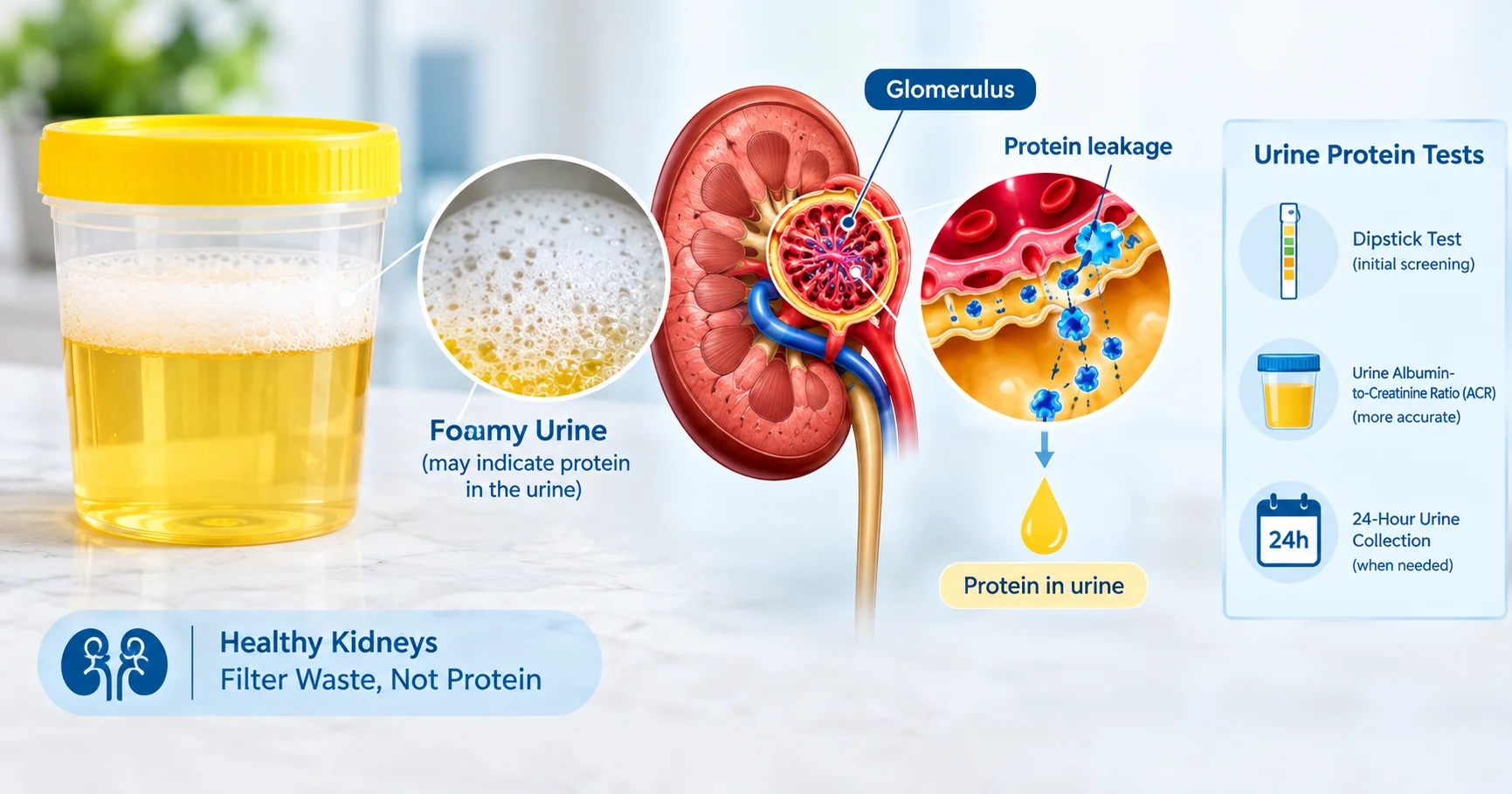
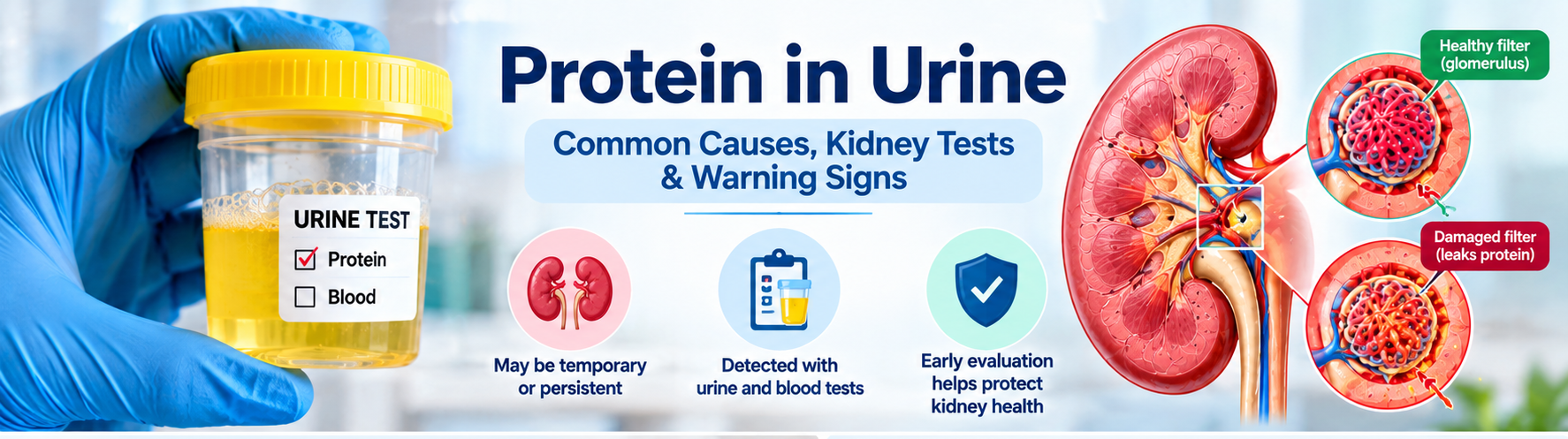
Short answer : Protein in urine can be a fleeting finding after fever, intense exercise, or dehydration, but persistent proteinuria often signals damage to the kidney’s filtering units (glomeruli). Ongoing leakage may require further testing and specialist care. Proteinuria—often discovered on a routine urine test—doesn’t always mean serious disease. A single abnormal result may be temporary, yet repeated or high‑level protein loss usually points to kidney‑filter damage that needs evaluation. What Is Proteinuria? Healthy kidneys keep large proteins such as albumin inside the bloodstream while allowing waste and excess fluid to pass into urine. The glomeruli act as tiny sieves; when they are inflamed, scarred, or otherwise damaged, proteins slip through and appear in the urine. How the kidneys normally work  – Blood enters the glomerulus at high pressure.  – Small molecules (water, electrolytes, waste) filter out. – Large proteins are retained and re‑absorbed in the tubules. When the glomerular barrier is compromised, albumin and other proteins are lost, leading to a measurable proteinuria. Common Causes of Protein in Urine (Persistent) These conditions often produce ongoing protein leakage and merit a thorough work‑up:  – Diabetes mellitus – high blood sugar damages glomerular capillaries.  – Hypertension (high blood pressure) – pressure overload injures the filtration barrier.  – Glomerular diseases – e.g., IgA nephropathy, membranous nephropathy, focal segmental glomerulosclerosis.  – Chronic infections – hepatitis C, HIV, or recurrent urinary‑tract infections.  – Cardiovascular conditions – heart failure can raise kidney venous pressure.  – Medications – non‑steroidal anti‑inflammatory drugs (NSAIDs), certain antibiotics, and some chemotherapy agents.  – Other kidney disorders – polycystic kidney disease, obstructive uropathy. For an in‑depth look at these factors, see Causes of Protein in Urine: Understanding the Key Factors. Temporary Causes of Protein in Urine Not every positive protein test signals chronic disease. Transient proteinuria often resolves once the trigger disappears: Fever or acute illness – inflammation raises glomerular permeability. Strenuous exercise – muscle breakdown and increased blood flow can spill protein. Dehydration – concentrated urine may falsely appear protein‑rich. Cold exposure – vasoconstriction can temporarily affect filtration. Recent urinary‑tract infection – inflammation of the bladder or urethra. Because a single abnormal result may be fleeting, clinicians often repeat a morning urine sample before ordering extensive work‑up. Symptoms and Warning Signs Early proteinuria is frequently silent, but watch for these clues:  – Foamy, frothy, or bubbly urine.  – Swelling (edema) of ankles, feet, or around the eyes.  – Unexplained fatigue or loss of appetite.  – Changes in urine volume or frequency.  – Elevated blood pressure. Most people with mild proteinuria feel fine; the presence of symptoms often indicates a higher level of protein loss or concurrent kidney dysfunction. Does foamy urine always mean protein in urine? No. Foam can result from rapid urine flow, concentrated urine, or even certain detergents in the toilet bowl. Only a laboratory test (dipstick or ACR) can confirm proteinuria. Protein in Urine vs. Blood in Urine Proteinuria (protein) and hematuria (blood) are distinct findings. Protein indicates a leak of albumin or larger proteins, while blood signals red‑cell leakage, often from a different part of the urinary tract. When both appear together, clinicians consider glomerular disease or severe inflammation. Read more about the overlap in our protein and blood in urine article. How Proteinuria Is Measured The most common tests include: – Urine dipstick – quick screening, semi‑quantitative.  – Urine albumin‑to‑creatinine ratio (UACR) – expresses albumin in mg per gram of creatinine; preferred for diabetes monitoring.  – Urine protein‑to‑creatinine ratio (PCR) – measures total protein.  – Comprehensive urinalysis – evaluates sediment, cells, and casts.  – Serum creatinine & eGFR – assess overall kidney filtration function. Interpretation of UACR:Range (mg/g)Interpretation<30Normal or low‑grade30–300Micro‑albuminuria (early kidney damage)>300Macro‑albuminuria (significant kidney disease) Urine ACR (UACR) Result Interpretation UACR Result Interpretation Below 30 mg/g Normal to mildly increased 30–300 mg/g Moderately increased albuminuria Above 300 mg/g Severely increased albuminuria Results should be repeated, especially if the first sample was a random collection, and interpreted alongside creatinine, eGFR, and clinical context. Can You Have Protein in Urine with Normal Creatinine? Yes. Protein leakage can precede a rise in serum creatinine because creatinine reflects overall filtration, not the selective loss of albumin. Early glomerular injury may show a high UACR while eGFR remains >90 mL/min/1.73 m². Glomerular Disease: When to Be Concerned Persistent, significant proteinuria—especially when accompanied by any of the following—should raise suspicion for a primary glomerular disorder:  – Protein levels >300 mg/g (macro‑albuminuria).  – Concurrent hematuria (blood in urine).  – Unexplained edema or high blood pressure.  – Declining eGFR over months.  – Abnormal urine sediment (casts, dysmorphic RBCs). Dr. Jyoti Bansode has a special clinical interest in glomerular diseases; patients with these red flags are often referred for nephrology evaluation. When Might a Kidney Biopsy Be Needed? Most proteinuria cases are managed without invasive procedures. A biopsy is considered when:  – The underlying cause remains unclear after standard labs and imaging.  – Protein loss exceeds 3.5 g/day (nephrotic‑range) with rapid eGFR decline.  – There is suspicion of a specific glomerular disease that guides therapy (e.g., lupus nephritis, IgA nephropathy).  – Patients have refractory proteinuria despite optimal blood‑pressure and diabetes control. Read more about the procedure in our kidney biopsy overview. Patient Checklist After an Abnormal Urine Protein Test Ask which test was performed (dipstick, UACR, PCR). Confirm the exact protein value and its units. Request your serum creatinine and eGFR results. Discuss any recent fever, intense exercise, dehydration, or infection that could be temporary. Inquire whether a repeat morning urine sample is recommended. Review blood‑pressure control and diabetes management. Ask if a referral to a nephrologist is appropriate. When to See a Nephrologist Consider specialist care if you have any of the following:  – Persistent proteinuria on two separate tests (≥30 mg/g).  – Rising UACR or PCR over weeks to months.  – Proteinuria accompanied by hematuria.  – Declining eGFR (<60 mL/min/1.73 m²) or rising serum creatinine.  – Significant edema or uncontrolled hypertension.  – Suspected glomerular disease based on urine sediment.  – Unclear cause after standard work‑up. Schedule a consultation through our appointment and consultation page. Medical ReviewDr. Jyoti
Kidney Tests Explained: Creatinine, eGFR, Urine ACR & Electrolytes

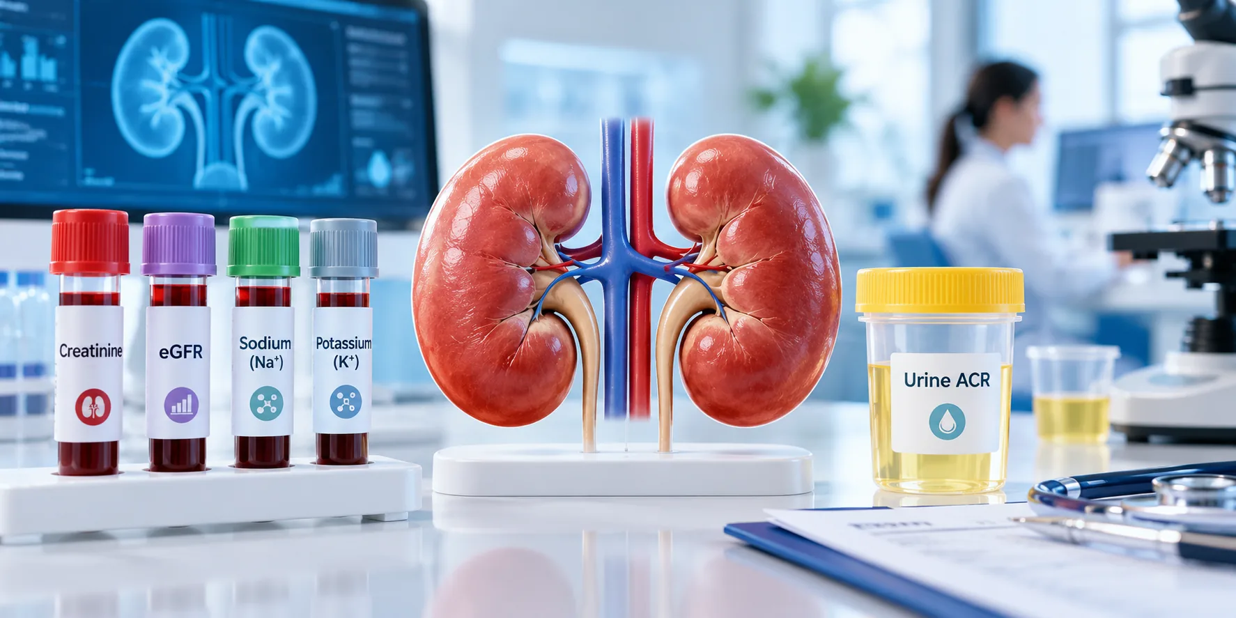
Kidney Tests Explained: Creatinine, eGFR, Urine ACR & ElectrolytesUnderstanding kidney function starts with the right lab tests. Creatinine, eGFR, urine albumin‑to‑creatinine ratio (uACR) and electrolyte panels are the four pillars doctors use to spot early disease, track progression, and guide treatment. This guide breaks down each test, normal values, and what the numbers mean for you. Medical Review – Dr. Jyoti BansodeMBBS, MD Medicine, DM NephrologyOver 10 years of experience in diagnosing and managing kidney disease. For personalized care, visit Dr. Bansode’s nephrology services. Why Kidney Function Testing Matters Kidneys filter waste, balance fluids, and regulate electrolytes. When they falter, waste builds up, blood pressure spikes, and vital minerals become imbalanced. Early detection through lab testing can prevent irreversible damage and reduce the need for dialysis or transplant. Common Symptoms That Prompt Testing Persistent fatigue or weakness Swelling in ankles, feet, or face Changes in urine color or frequency Unexplained high blood pressure Muscle cramps or tingling sensations If you notice any of these, ask your primary‑care physician for a kidney panel. Key Kidney Tests Overview Key Kidney Tests Overview Serum Creatinine – What It Shows Creatinine is a waste product from muscle metabolism. The kidneys clear it from the blood, so higher levels indicate reduced filtration. Normal ranges vary by sex and muscle mass, but typical limits are 0.6–1.2 mg/dL for women and 0.7–1.3 mg/dL for men Nephrology Specialists. Values above these thresholds often signal early kidney stress. Estimated Glomerular Filtration Rate (eGFR) eGFR translates the creatinine value into an estimate of how many milliliters of blood the kidneys filter each minute per 1.73 m² of body surface. An eGFR > 60 mL/min/1.73 m² is generally considered normal, 30–59 suggests moderate decline, and <30 indicates severe kidney disease MyNephrologist. The calculation also incorporates age, sex, and sometimes race, making it a more comprehensive indicator than creatinine alone. Urine Albumin‑to‑Creatinine Ratio (uACR) Albumin is a protein that should stay in the bloodstream. When the glomeruli are damaged, albumin leaks into urine. uACR compares the amount of albumin to creatinine in a single urine sample, eliminating the need for a 24‑hour collection. Normal uACR is 300 mg/g reflects macro‑albuminuria (significant damage) MyNephrologist. This test can detect kidney injury years before eGFR drops. Electrolytes and Blood Urea Nitrogen (BUN) Electrolyte panels measure sodium, potassium, chloride, bicarbonate, and calcium—minerals the kidneys tightly regulate. Abnormalities often accompany chronic kidney disease (CKD). BUN, another waste product, rises as kidney function declines but is less specific than creatinine because it is influenced by diet and hydration Nephrology Specialists. Interpreting Results Together Creatinine & eGFR Relationship Because eGFR is derived from creatinine, the two numbers should be read as a pair. A mildly elevated creatinine with a normal eGFR may be due to high muscle mass, while a normal creatinine with a low eGFR often points to early CKD, especially in older adults. When uACR Raises a Red Flag Even with a healthy eGFR, a uACR > 30 mg/g signals that the kidney’s filtering units are leaking protein. This early sign is common in diabetes and hypertension and warrants tighter blood‑sugar or blood‑pressure control, plus possible referral to a nephrologist. Electrolyte Imbalance Clues High potassium (hyperkalaemia) can indicate reduced excretion, while low sodium often reflects fluid overload. Metabolic acidosis (low bicarbonate) is another hallmark of advanced CKD. Spotting these changes early allows dietary or medication adjustments before complications arise. When to Seek Specialist Care If any of the following appear, schedule a consultation with a kidney specialist: eGFR consistently below 60 mL/min/1.73 m² uACR ≥ 30 mg/g on two separate tests Persistent electrolyte abnormalities despite treatment Rapid rise in serum creatinine (e.g., >0.3 mg/dL in 48 hours) Our clinic’s nephrology team can provide a detailed assessment and personalized plan. Learn more about when to see a specialist in When Should You See a Nephrologist in Navi Mumbai?. Common Myths About Kidney Tests Myth: “If my creatinine is normal, my kidneys are fine.” – Fact: Creatinine alone can miss early damage; uACR often reveals problems first. Myth: “Only diabetics need kidney testing.” – Fact: Hypertension, family history, and age over 50 are also risk factors. Myth: “Low potassium always means a kidney problem.” – Fact: Diet, medications, and gastrointestinal loss can also lower potassium. Putting It All Together: A Practical Checklist Use this quick reference during your next lab visit: Kidney Test Typical / Reference Range When to Seek Medical Attention Serum Creatinine Reference ranges vary by laboratory, age, sex, and muscle mass. A persistently elevated or rising creatinine level may require further evaluation. eGFR Generally ≥60 mL/min/1.73 m² An eGFR below 60 that persists for 3 months or longer may indicate chronic kidney disease. Urine ACR (uACR) <30 mg/g 30–300 mg/g indicates moderately increased albuminuria; >300 mg/g indicates severely increased albuminuria. Electrolytes(Na, K, Cl, HCO₃) Within your laboratory’s reference range Persistent or significant abnormalities, particularly potassium or bicarbonate changes, require medical assessment. BUN Typically about 7–20 mg/dL, depending on the laboratory An elevated BUN should be interpreted together with creatinine, eGFR, hydration status, and other clinical findings. Regular monitoring—typically every 6–12 months for at‑risk patients—helps catch changes early. For detailed guidance on test frequency, see our When Is Dialysis Needed? Symptoms, eGFR & Clinical Factors page. By understanding what each number represents, you can partner with your healthcare team to protect kidney health before problems become serious. Frequently Asked Questions What should patients know first about kidney disease tests? Kidney tests measure waste removal (creatinine, eGFR), protein leakage (uACR), and mineral balance (electrolytes). Together they reveal how well the kidneys filter blood and whether early damage is present.   Which kidney tests are most relevant for early detection? The urine albumin‑to‑creatinine ratio (uACR) is the most sensitive early marker, often detecting damage before eGFR declines. Serum creatinine and eGFR confirm overall filtration capacity.   What symptoms should prompt evaluation? Persistent fatigue, swelling, changes in urine, high blood pressure, and unexplained muscle cramps should trigger a kidney panel, including creatinine, eGFR, uACR, and electrolytes.   How are creatinine and eGFR interpreted together? Creatinine provides the raw waste level, while eGFR translates it into an estimated filtration

How to Prevent Kidney Stones Recurrence – Proven Steps

Kidney stones can be excruciating, and the fear of another episode often looms large after the first pass. Fortunately, research shows that simple, consistent habits can cut the risk of recurrence dramatically. Below you’ll find evidence‑based strategies, the tests that guide personalized care, and clear guidance on when to involve a kidney specialist. Why Preventing Recurrence Matters Each stone event can cause severe pain, time off work, and costly medical visits. Repeated stones also increase the risk of chronic kidney damage and may lead to surgical interventions. Proactive prevention therefore protects both health and quality of life. Who May Be at Higher Risk? Risk factors include a history of stones, certain metabolic conditions (e.g., hypercalciuria, gout), a diet high in sodium or animal protein, low fluid intake, and a family history of nephrolithiasis. People with obesity, diabetes, or a previous urinary tract infection also face higher odds of recurrence. Practical Daily Prevention Steps. Daily habits to prevent kidney stones including hydration, healthy diet, reduced salt and regular follow-up Hydration – The Cornerstone Most guidelines recommend producing 2.5–3 L of urine per day, which typically means drinking about 3 L (5.2 pints) of fluid daily. Water is best, but tea, coffee, and low‑sugar fruit juices also count. Adding fresh lemon juice can increase urinary citrate, a natural stone inhibitor. Carry a reusable bottle and sip continuously. Set hourly reminders on your phone. Track urine volume; aim for a pale yellow color. Dietary Adjustments Tailor your diet to the type of stone you’ve formed. General recommendations, supported by NYU Langone Health, include: Limit animal protein to 6–8 oz per day (about two decks of cards). Reduce sodium to < 2,300 mg daily; aim for 1,500 mg if you’re salt‑sensitive. Increase fruits and vegetables, especially those high in citrate (citrus, berries, kiwi). Choose low‑oxalate foods if you have calcium‑oxalate stones (e.g., limit spinach, nuts, and chocolate). Lifestyle Considerations Beyond food and fluid, these habits matter: Maintain a healthy weight; obesity raises calcium and uric acid excretion. Exercise regularly – moderate activity improves calcium metabolism. Avoid excessive sugary drinks and carbonated beverages, which increase urinary calcium and uric acid. Limit vitamin C supplements above 1 g/day, as excess converts to oxalate. Common Mistakes to Avoid Many patients unintentionally increase risk: Relying on coffee alone for hydration – caffeine can be mildly diuretic. Thinking “low‑calcium” diets prevent stones – inadequate calcium can raise oxalate absorption. Skipping the 24‑hour urine test – without objective data, dietary tweaks may miss the mark. When Individualized Advice Is Needed If you’ve had more than one stone, a metabolic evaluation is essential. A 24‑hour urine stone test measures volume, pH, calcium, oxalate, citrate, uric acid, and sodium, helping clinicians tailor prevention. 24-hour urine test for recurrent kidney stones showing urine collection, risk factors and prevention steps Action What to Do What to Limit / Avoid When to Discuss With Doctor 💧 Fluid Intake Drink about 3 L of fluid daily Sugary sodas and excess soft drinks If urine output remains low despite adequate fluid intake 🧂 Salt Keep sodium below 2,300 mg/day unless advised otherwise Processed foods and salty snacks If blood pressure or urinary sodium is high 🍗 Animal Protein Keep portions moderate Excess red meat and processed meat If uric acid is high on a 24-hour urine test 🥛 Calcium Get adequate calcium from food Unnecessary high-dose calcium supplements If calcium-based kidney stones keep recurring In a concise prevention statement: Stay well‑hydrated, limit salt and animal protein, eat plenty of fruits/vegetables, and have a 24‑hour urine test to fine‑tune your plan. Medical Review Dr. Jyoti Bansode, MBBS, MD Medicine, DM Nephrology – Over 10 years of nephrology experience. For personalized guidance, visit our Kidney Stone Preventions page or schedule a consultation through our When Should You See a Nephrologist service. For deeper reading, see the NHS prevention guide NHS guidance and the AAFP lifestyle recommendations AAFP article. Frequently Asked Questions How much water should I drink to stop kidney stones from returning? Aim for 2.5–3 L of urine output daily, which usually means drinking about 3 L (5 pints) of fluid spread throughout the day. Can a low‑calcium diet prevent kidney stones? No. Adequate dietary calcium (1,000–1,200 mg) binds oxalate in the gut, reducing stone risk; only high‑dose supplements should be limited. What does a 24‑hour urine stone test reveal? It measures volume, pH, calcium, oxalate, citrate, uric acid, and sodium, allowing doctors to customize fluid, diet, and medication plans. When should I see a nephrologist instead of a urologist? If you have recurrent stones, abnormal urine test results, or metabolic conditions (e.g., hyperparathyroidism), a nephrologist can manage the underlying cause. Does adding lemon to water help? Yes. Lemon juice raises urinary citrate, which binds calcium and helps prevent calcium‑oxalate stone formation. Conclusion Preventing kidney stone recurrence is within reach by drinking enough water, adjusting your diet, and using targeted testing. Start these habits today and consult a nephrologist if you notice any warning signs.

Kidney Specialist in Vashi: What to Bring to Your Consultation

Visiting a kidney specialist (nephrologist) for the first time can feel overwhelming, especially if you’re unsure which documents or test results to carry. In Vashi, NewEra Hospitals hosts Dr. Jyoti Bansode, a seasoned nephrologist with more than a decade of experience. This guide walks you through the exact items you should prepare, the symptoms that prompt a visit, and how the consultation typically unfolds, so you can feel confident and ready. When Should You See a Kidney Specialist in Vashi? Kidney health often goes unnoticed until a problem becomes evident through lab values or symptoms. According to When Should You See a Nephrologist in Navi Mumbai?, you should consider a referral when any of the following occur: Persistent swelling in the ankles, feet, or face. Unexplained fatigue or weakness despite adequate rest. Changes in urine colour, frequency, or the presence of blood. High blood pressure that is difficult to control with medication. Elevated serum creatinine or a declining eGFR on routine blood work. These red‑flag signs often indicate that a detailed renal evaluation is needed. Common Symptoms That Prompt a Nephrology Referral While some kidney issues are detected incidentally, many patients notice physical cues. Typical symptoms include: Foamy or bubbly urine – a sign of proteinuria. Persistent lower‑back or flank pain, which may suggest stones or infection. Frequent urination at night (nocturia). Unexplained itching, especially on the arms and legs. Loss of appetite or nausea. If you experience any of these, schedule an appointment promptly. Nephrology consultation checklist showing lab reports, kidney ultrasound, medications, medical history and questions to ask What to Bring to Your Consultation Arriving prepared saves time and ensures the nephrologist can make an accurate assessment. Below is a comprehensive checklist. Essential Lab Reports Blood and urine tests form the backbone of kidney evaluation. Bring the most recent reports, ideally from the past 3 months: Serum Creatinine & eGFR – shows current filtration rate. Blood Urea Nitrogen (BUN) – complements creatinine. Electrolytes (Na+, K+, Cl-, HCO3-) – reveals imbalances. Urine Albumin‑to‑Creatinine Ratio (UACR) or 24‑hour urine protein – quantifies protein loss. Lipid profile – important for cardiovascular risk in CKD patients. These tests are highlighted in What to Expect at Your First Visit with a Nephrologist, which notes that most doctors request a full panel before the first appointment. Imaging and Other Diagnostic Documents If you have already undergone imaging, bring copies of the reports: Renal ultrasound – assesses kidney size and structural abnormalities. CT or MRI scans – useful for complex stone disease or tumors. Previous kidney biopsies – include pathology results if available. Even if the imaging was performed at another facility, a printed PDF or CD will help Dr. Bansode review the findings without delay. Personal Health History Prepare a concise summary of your medical background: List of current medications, including over‑the‑counter supplements. History of diabetes, hypertension, or autoimmune diseases. Family history of kidney disease or transplant. Any prior surgeries or hospitalizations related to the urinary tract. Having this information on hand enables the doctor to spot patterns and tailor treatment. How Dr. Jyoti Bansode Evaluates Your Case Dr. Jyoti BansodeMBBS, MD Medicine, DM Nephrology10+ years of nephrology experiencePractice at NewEra Hospitals, Vashi Dr. Bansode follows a systematic approach that blends clinical expertise with evidence‑based guidelines. Review of Tests and Imaging During the appointment, the nephrologist will first examine your lab values and imaging reports. Patterns such as a steadily falling eGFR or persistent proteinuria guide the next steps—whether it’s a medication adjustment, a repeat test, or a referral for a kidney biopsy. Physical Examination The doctor will assess blood pressure (both sitting and standing), check for edema, and listen for heart or lung sounds that may indicate fluid overload. A focused abdominal exam helps identify kidney enlargement or tenderness. Discussion & Plan After the assessment, Dr. Bansode will explain the diagnosis in plain language, outline lifestyle modifications, and prescribe any necessary medications. She also discusses the need for follow‑up labs, potential referrals (e.g., dietitian, transplant team), and answers any questions you have. Checklist for Your Appointment What to bring to a nephrology consultation including kidney test reports, imaging, medications and medical history Symptoms to Mention: swelling, foamy urine, nocturia, fatigue, itching. Lab Reports: serum creatinine, eGFR, BUN, electrolytes, urine protein, lipid profile. Imaging: renal ultrasound, CT/MRI, biopsy reports (if any). Medication List: include doses, frequency, and any herbal supplements. Health History: diabetes, hypertension, family kidney disease, past surgeries. Questions to Ask: What does my eGFR mean for daily life? Are there diet changes I should adopt now? Do I need a repeat ultrasound or a kidney biopsy? What are the signs that require urgent care? Urgent Care Triggers: sudden drop in urine output, severe flank pain, rapid rise in blood pressure, swelling of the face or lungs. Preparing for a Second Opinion If you’ve already seen another nephrologist and want a second perspective, bring the same set of documents listed above, plus any written treatment plans or referral letters. Dr. Bansode can compare the previous recommendations with her own assessment, ensuring you receive the most comprehensive care. When to Seek Urgent Care in Vashi Kidney emergencies require immediate attention. According to Nephrology Services in Vashi | MGM New Bombay Hospital, the following situations merit a 24‑hour visit or a call to emergency services: Sudden, severe flank pain that radiates to the groin (possible kidney stone blockage). Rapid swelling of the legs, abdomen, or face accompanied by shortness of breath. Marked increase in blood pressure (>180/110 mmHg) that does not respond to medication. Decreased urine output (<400 ml per day) or complete anuria. Fever with chills and urinary symptoms, suggesting a kidney infection. In these cases, head straight to the emergency department at NewEra Hospitals or call the local emergency number. By gathering the right paperwork and understanding what to expect, you empower Dr. Bansode to deliver focused, effective kidney care. For appointment scheduling, visit the Best Kidney Specialist in Vashi – Dr. Rashmi Yadav directory to see how other specialists in the area

When Should You See a Nephrologist in Navi Mumbai?

When Should You See a Nephrologist in Navi Mumbai? Kidney health often goes unnoticed until a problem becomes serious. Knowing the right moment to consult a kidney specialist can prevent complications, preserve function, and improve quality of life. This guide explains the warning signs, essential lab reports, and what to expect during a nephrology visit in Navi Mumbai, with a focus on the expertise of Dr. Jyoti Bansode. Why Early Nephrology Care Matters Kidneys filter waste, balance electrolytes, and regulate blood pressure. When they start to falter, the body may not show obvious symptoms right away. Early detection—especially for people with diabetes, hypertension, or a family history of kidney disease—allows timely interventions that can slow or even reverse damage. According to Prime Care 360’s kidney care guide, many kidney conditions are silent in the first stages, making regular monitoring essential. Key Situations When You Should See a Nephrologist Abnormal Blood Tests: Elevated serum creatinine, low eGFR (<60 mL/min/1.73 m²), or rising blood urea nitrogen (BUN) without an obvious cause. Urine Changes: Persistent proteinuria, blood in urine, foamy or frothy urine, or frequent nighttime urination. Swelling (Edema): Unexplained swelling in the ankles, feet, face, or hands. High Blood Pressure Uncontrolled by Medication: Hypertension that remains high despite treatment can be a sign of kidney involvement. Chronic Conditions: Diabetes, heart disease, or autoimmune disorders (e.g., lupus) that increase kidney risk. Family History: A parent or sibling with chronic kidney disease (CKD) or kidney failure. If any of these red flags appear, schedule a kidney consultation promptly. Essential Reports to Bring to Your Appointment Having the right documentation helps the nephrologist assess your kidney health efficiently. Prepare the following: Recent serum creatinine and eGFR results. Urine analysis reports (protein, blood, microalbumin). Blood pressure logs (if you monitor at home). Medication list, including over‑the‑counter supplements. Any prior imaging (ultrasound, CT) or biopsy reports. For a deeper dive into interpreting eGFR, see our What Does Your eGFR Number Mean? article. How Dr. Jyoti Bansode Evaluates Kidney Problems Medical Review – Dr. Jyoti Bansode MBBS, MD (Medicine), DM (Nephrology) – over 10 years of dedicated nephrology practice. Based in NewEra Hospitals, Vashi, Dr. Bansode offers comprehensive kidney care, from early CKD detection to dialysis planning and transplant evaluation. Patients benefit from a personalized approach, state‑of‑the‑art diagnostics, and coordinated care with allied specialists. During the first visit, Dr. Bansode typically: Reviews your medical history and risk factors. Analyzes the lab reports you bring. Performs a focused physical exam (checking for edema, blood pressure, and abdominal masses). Orders additional tests if needed (e.g., renal ultrasound, 24‑hour urine protein, or kidney biopsy). Creates a tailored management plan, which may include lifestyle changes, medication adjustments, or referral for advanced therapies. Questions to Ask at Your Nephrology Consultation Being proactive with questions ensures you leave the appointment with a clear roadmap. Consider asking: What is the likely cause of my kidney abnormalities? How fast is my kidney function declining, and what does my eGFR trend mean? Which lifestyle changes can protect my kidneys the most? Do I need medication adjustments for blood pressure or diabetes? When should I return for follow‑up labs or imaging? Is a second opinion or referral to a transplant center appropriate? When Kidney Symptoms Require Urgent Attention Kidney warning signs that require urgent medical attention including pain, swelling, fever, low urine output and breathlessness Some kidney‑related emergencies need immediate medical care. Seek urgent evaluation if you experience: Sudden, severe flank pain or blood in the urine. Rapid swelling of the face or legs accompanied by shortness of breath. Marked drop in urine output (oliguria) or complete absence of urine (anuria). High fever with chills and flank tenderness (possible kidney infection). Confusion, nausea, or vomiting with rising creatinine levels. In such cases, go to the nearest emergency department or call emergency services. Preparing for Your Nephrologist Appointment To make the most of your visit: Fast for 8‑12 hours if you’re scheduled for blood work. Bring a list of all medications, including herbal supplements. Write down any symptoms, their frequency, and triggers. Carry previous kidney‑related reports (e.g., What Causes High Creatinine? for background). Prepare insurance or payment information; most clinics in Navi Mumbai accept major insurers. Finding a Trusted Kidney Specialist in Navi Mumbai Beyond Dr. Jyoti Bansode, Navi Mumbai hosts several reputable nephrology centers, such as those listed on Practo’s nephrologist guide and Echelon Hospital’s nephrology services. When choosing a provider, consider credentials, patient reviews, and proximity to your home or workplace. Consistency in follow‑up care is key for chronic kidney disease management. Local Support and Resources Living in Navi Mumbai offers access to specialized kidney care facilities, patient education workshops, and support groups. Organizations often host free screening camps for diabetes and hypertension—two major contributors to kidney disease. Staying informed and participating in community health events can help you catch problems early. Frequently Asked Questions When should I schedule my first nephrology appointment? Book a visit as soon as you have abnormal kidney labs (e.g., high creatinine or low eGFR), persistent proteinuria, unexplained swelling, or uncontrolled high blood pressure. What lab tests are most important for a kidney evaluation? Serum creatinine, eGFR, BUN, urine protein/albumin, and a complete metabolic panel are essential; a 24‑hour urine protein test may be added if needed. Can I get a second opinion on a kidney diagnosis? Yes, seeking a second opinion is encouraged, especially before major decisions like dialysis initiation or transplant evaluation. How often should I have kidney function checked if I have diabetes? People with diabetes should have kidney function tests (creatinine, eGFR, and urine albumin) at least twice a year, or more frequently if abnormalities are detected. What lifestyle changes help preserve kidney health? Maintain blood pressure and blood sugar control, stay hydrated, limit excessive salt and protein, avoid NSAIDs overuse, and quit smoking. When is kidney disease considered an emergency? Sudden severe flank pain, rapid swelling, sudden loss of urine output, high fever with flank tenderness, or confusion with rising creatinine require immediate medical attention. Conclusion Protecting your kidneys starts with recognizing the signs

Kidney Transplant Evaluation: When Should Preparation Begin?

Facing kidney failure can feel overwhelming, but understanding the transplant evaluation process empowers you to act at the right moment. This guide explains when to start preparing, what the evaluation entails, and how to navigate each step with confidence. Why Kidney Transplant Evaluation May Be Discussed A transplant nephrologist typically raises the topic when your kidney function declines to a level where dialysis or transplant becomes the most viable long‑term solution. According to the National Kidney Foundation, patients are usually referred once their glomerular filtration rate (GFR) falls between 25 and 30 mL/min/1.73 m². The HRSA guidelines echo this, noting that early referral gives the transplant team ample time to complete testing before end‑stage renal disease (ESRD) sets in. Who May Need an Evaluation? Not every person with chronic kidney disease (CKD) qualifies for a transplant right away. Candidates typically include: Adults with CKD stage 4 (eGFR 15‑29) or stage 5 (eGFR < 15) who are otherwise medically stable. Patients already on dialysis who meet age, cardiovascular, and psychosocial criteria. Living donors – family members or friends with compatible blood type and kidney health. High‑risk or ABO‑incompatible cases managed by experienced centers like Dr. Jyoti Bansode’s practice. Understanding your eGFR is crucial; see our eGFR guide for a detailed breakdown. How to Prepare for the Evaluation How to Prepare for the Evaluation Preparation starts well before the first appointment. Gather the following: Complete medical history, including past surgeries, infections, and vaccination records. Recent laboratory results (CBC, metabolic panel, hepatitis B/C, HIV, and blood type). Imaging reports (chest X‑ray, abdominal ultrasound, cardiac stress test if indicated). List of current medications, especially immunosuppressants or anticoagulants. Psychosocial documentation – proof of stable housing, insurance, and support system. Below is a quick reference table that outlines what to do before, during, and after the evaluation. Phase Key Actions Purpose Before Collect records, schedule labs, arrange transportation Ensure the transplant center has a complete picture During Undergo physical exam, blood work, imaging, and psychosocial interview Assess medical suitability and match potential After Review results with the transplant team, discuss listing status Determine next steps – wait‑list, additional testing, or alternative therapy What Happens During the Evaluation The transplant center conducts a comprehensive, one‑day assessment that may include: Blood tests: kidney function panel, HLA typing, infectious disease screening. Imaging: Doppler ultrasound of kidneys, chest X‑ray, and possibly a cardiac echo. Cardiovascular review: stress test or coronary calcium scan for patients over 50 or with heart risk factors. Psychosocial interview: evaluation of mental health, adherence potential, and support network. Educational session: overview of post‑transplant care, medication regimen, and lifestyle changes. For a detailed look at the testing protocol, refer to the UPMC evaluation overview. Recovery, Results, and Follow‑Up Most patients leave the evaluation center the same day, but the decision timeline varies. Centers typically provide a decision within 1‑2 weeks. If approved, you’ll be placed on the national waiting list and matched based on HLA compatibility, blood type, and waiting time. Post‑evaluation follow‑up includes: Review of any abnormal findings (e.g., cardiac issues that need treatment before listing). Vaccination updates – especially hepatitis B and pneumococcal vaccines. Continued dialysis planning if you’re already on treatment. Regular meetings with the transplant coordinator to keep your profile current. Warning signs of kidney problems that need immediate medical review Warning Signs That Need Immediate Medical Review Even after a successful evaluation, certain symptoms warrant prompt attention: Sudden swelling of the legs or face. Fever, chills, or unexplained infections. Shortness of breath or chest pain. New onset of severe hypertension. Rapid decline in urine output (if you still produce urine). Report any of these to your nephrologist right away to avoid complications. Medical Review – Dr. Jyoti Bansode Dr. Jyoti Bansode holds an MBBS, MD in Medicine, and a DM in Nephrology, with over 10 years of experience. She has performed high‑risk, ABO‑incompatible, and pediatric kidney transplants, and leads a dedicated transplant team in Navi Mumbai. For personalized guidance, schedule an dialysis consultation or contact her office directly. Preparing early, understanding each step, and staying vigilant about warning signs can make the difference between a smooth transplant journey and unnecessary delays. Frequently Asked Questions When is the ideal time to begin a kidney transplant evaluation? Experts recommend starting the evaluation when the eGFR falls between 25 and 30 mL/min/1.73 m², allowing enough time to complete testing before reaching end‑stage renal disease. What tests are required during the transplant evaluation? The evaluation typically includes blood work for kidney function and HLA typing, imaging such as chest X‑ray and kidney ultrasound, cardiac screening, and a psychosocial interview. Can I be evaluated for a transplant while still on dialysis? Yes. Patients on dialysis are often evaluated concurrently so they can be listed immediately if they meet all medical and psychosocial criteria. How long does it take to receive the evaluation results? Most transplant centers provide a decision within 1–2 weeks after the one‑day assessment, though complex cases may require additional testing. What should I do if I develop a fever after the evaluation? Fever can signal infection, which is a red‑flag for transplant candidates. Contact your nephrologist or transplant coordinator immediately. Are there special considerations for living donors? Living donors undergo a parallel evaluation to ensure kidney health, blood type compatibility, and overall suitability, often completing the process faster than deceased‑donor pathways. Conclusion Start the evaluation process as soon as your eGFR approaches 25 mL/min/1.73 m², gather all required documents, and keep an open line of communication with your transplant team. Early preparation maximizes your chances of a timely, successful kidney transplant.

Kidney Biopsy Preparation & Recovery: What to Expect

A kidney biopsy provides essential clues about kidney health, but the thought of a needle in your back can be intimidating. This guide walks you through why doctors order the test, how to get ready, what the procedure looks like, and the steps for a safe, comfortable recovery. Why a Nephrologist May Recommend a Kidney Biopsy Nephrologists use kidney biopsies to pinpoint the exact cause of kidney problems when blood tests, urine studies, or imaging cannot give a clear answer. The tissue sample helps differentiate between glomerular diseases, interstitial nephritis, vascular disorders, and other conditions that require specific treatment. Who Might Need a Biopsy? Unexplained proteinuria or hematuria that persists despite treatment. Rapidly declining kidney function (eGFR drop) without an obvious cause. Suspected glomerular disease such as IgA nephropathy or lupus nephritis. Evaluation of a kidney mass or cyst before surgery. Monitoring response to therapy in known kidney disease. Understanding the reason behind the test helps you feel more in control and can guide post‑procedure discussions with your doctor. Preparing for Your Kidney Biopsy Preparation is mostly about safety and comfort. Follow these steps to minimize complications: Medication review: Stop blood‑thinners (aspirin, warfarin, clopidogrel) and non‑steroidal anti‑inflammatory drugs (NSAIDs) as instructed, usually 5–7 days before the procedure. Fasting: Do not eat or drink after midnight if you will receive sedation. Lab checks: Your doctor will order a complete blood count, coagulation profile, and basic metabolic panel to ensure you are ready. Bring a support person: You will need someone to drive you home and stay with you for the first 24 hours. Write down questions: Note any concerns about pain, bleeding risk, or medication adjustments. According to Kidney Fund guidance, clear communication reduces anxiety and improves outcomes. For more on interpreting kidney function numbers, see our eGFR test article. What Happens During the Procedure The biopsy is usually performed under local anesthesia with real‑time ultrasound guidance. A thin needle is inserted through the flank into the kidney to obtain 2–3 core samples. The entire process typically lasts 30–45 minutes. Phase What You Experience Key Goal Before Positioned on your side, IV line placed, skin cleaned. Ensure sterility and optimal needle trajectory. During Local anesthetic applied; needle advances under ultrasound. Collect adequate tissue while minimizing trauma. After Needle removed, pressure applied, bandage placed. Prevent immediate bleeding and start monitoring. Immediate Recovery in the Clinic After the biopsy, you will lie flat for several hours while nurses monitor vital signs, blood pressure, pulse, and urine output. This “flat‑lying” period reduces the risk of bleeding from the puncture site. Recovery Timeline (First 24 Hours) 0–6 hours: Strict bed rest; avoid sitting or standing upright. 6–12 hours: Light walking to the bathroom only if cleared. 12–24 hours: If vital signs are stable and urine is clear, you may be discharged home. Typical discharge instructions include drinking plenty of fluids, avoiding heavy lifting (>10 kg), and watching for signs of bleeding. At‑Home Recovery: Days 1–14 Kidney biopsy recovery at home with rest, hydration, urine monitoring and warning signs Most patients feel mild soreness at the biopsy site for a few days. Follow these guidelines for a smooth healing process: Activity: Rest for the first 24 hours, then resume light walking. Avoid strenuous exercise or heavy lifting for at least two weeks post‑biopsy recovery guide. Diet: Maintain a balanced diet rich in protein and fluids to support tissue repair. Limit salty foods that could raise blood pressure. Urine monitoring: Check urine for blood. A small amount of pink‑tinged urine is normal for 24–48 hours, but bright red urine or clots require immediate attention. Pain control: Over‑the‑counter acetaminophen is usually sufficient. Avoid NSAIDs unless your doctor approves. If you notice any of the warning signs below, call your nephrologist right away. Warning Signs That Need Prompt Medical Review Severe flank pain or increasing soreness. Bright red urine, large clots, or a sudden drop in urine output. Fever above 38 °C (100.4 °F) or chills. Unexplained dizziness, rapid heartbeat, or a sudden drop in blood pressure. These symptoms could indicate bleeding, infection, or other complications that need early intervention NIDDK recovery information. Understanding Your Biopsy Results Pathology results usually arrive within 3–7 days. The report will describe the type of kidney disease, degree of inflammation or scarring, and any specific findings that guide treatment. Your nephrologist will discuss the implications and may adjust medications accordingly. For related information on protein and blood in urine, visit the Kaiser Permanente home‑care guide. Medical Review – Dr. Jyoti Bansode MBBS, MD Medicine, DM Nephrology Over 10 years of nephrology experience and more than 200 kidney biopsies performed. All information is based on verified clinical guidelines and peer‑reviewed sources. Frequently Asked Questions How long do I need to stay in the hospital after a kidney biopsy? Most patients are discharged the same day after a few hours of observation, but some may stay overnight if there are concerns about bleeding or blood pressure. Is it normal to see blood in my urine after the biopsy? A small amount of pink‑tinged urine for 24–48 hours is common. Bright red urine, clots, or a sudden increase in bleeding should be reported immediately. When can I resume normal exercise after a kidney biopsy? Light walking is allowed after the first day, but avoid heavy lifting and vigorous exercise for at least two weeks, or until your doctor gives clearance. What medications should I stop before the procedure? Blood thinners (aspirin, warfarin, clopidogrel) and NSAIDs are typically stopped 5–7 days prior, but always follow the specific instructions from your nephrologist. How are kidney biopsy results delivered? Results are usually available within 3–7 days and are discussed in a follow‑up appointment where your doctor explains the diagnosis and treatment plan. Conclusion Preparing well, following post‑procedure instructions, and staying alert to warning signs can make your kidney biopsy recovery smooth and uneventful. If you have any doubts, contact your nephrology team promptly.

When Is Dialysis Needed? Symptoms, eGFR & Clinical Factors

Quick answer (40‑60 words): Dialysis is usually started when eGFR falls below 15 mL/min/1.73 m² and the patient shows uremic symptoms such as nausea, severe itching, fluid overload, high potassium, or metabolic acidosis. Even with higher eGFR, urgent dialysis may be required for life‑threatening electrolyte or fluid problems. Common symptoms: nausea, vomiting, loss of appetite, weight loss, severe itching, fatigue, confusion. Key tests: eGFR, serum potassium, bicarbonate, blood urea nitrogen, chest X‑ray for fluid overload. When to see a doctor: any of the above symptoms, eGFR < 20, uncontrolled blood pressure, or rapid rise in creatinine. Medical Review: Dr. Jyoti Bansode, MBBS, MD Medicine, DM Nephrology, with over 10 years of nephrology experience and more than 500 tunneled/Permacath catheter procedures, reviewed this content. Dialysis is a life‑saving therapy that replaces the filtering function of kidneys when they can no longer remove waste, excess fluid, or maintain electrolyte balance. Deciding when to start dialysis involves a blend of laboratory numbers, patient‑reported symptoms, and overall clinical context. What Is Dialysis and When Is It Considered? Dialysis can be performed as hemodialysis (blood filtered through a machine) or peritoneal dialysis (fluid in the abdomen absorbs waste). According to Mayo Clinic, hemodialysis is typically recommended when eGFR is very low and patients experience symptoms such as nausea, vomiting, loss of appetite, weight loss, severe itching, tiredness, or confusion. It is also indicated for fluid overload, high potassium, or severe metabolic acidosis. Understanding eGFR and the Critical Thresholds Estimated Glomerular Filtration Rate (eGFR) is the most widely used metric to gauge kidney function. Normal adult eGFR is >90 mL/min/1.73 m². Chronic Kidney Disease (CKD) is staged based on eGFR: Stage 3: eGFR 30‑59 Stage 4: eGFR 15‑29 Stage 5 (End‑Stage Renal Disease): eGFR <15 Guidelines from the National Kidney Foundation suggest that patients with Stage 5 CKD (eGFR < 15) are evaluated for dialysis or transplantation. However, the decision is not based on eGFR alone; clinical signs often drive the timing. Symptoms That Signal the Need for Dialysis Symptoms and clinical factors that may indicate the need for dialysis, including low eGFR, fluid overload, high potassium, nausea, and metabolic acidosis Even with an eGFR slightly above 15, dialysis may be necessary if severe symptoms cannot be managed medically. Key warning signs include: Uremic symptoms: nausea, vomiting, loss of appetite, weight loss, severe itching, fatigue, confusion, or seizures. Fluid overload: swelling in legs, shortness of breath, pulmonary edema, or uncontrolled hypertension. Electrolyte disturbances: potassium >6.5 mmol/L, severe metabolic acidosis (bicarbonate <15 mmol/L). Pericardial effusion or uremic pericarditis: chest pain, muffled heart sounds. Rapid decline in kidney function: creatinine rising >0.3 mg/dL within 48 hours. MyNephrologist notes that emergency dialysis is often required when kidney function drops to 10‑15 % of normal and life‑threatening complications arise. Clinical Factors Beyond eGFR Several non‑laboratory factors influence the decision: Blood pressure control: refractory hypertension despite optimal medication may indicate the need for dialysis. Nutritional status: unintentional weight loss, protein‑energy wasting, or malnutrition. Cardiovascular health: heart failure exacerbated by fluid overload. Patient preferences and quality of life: ability to adhere to dialysis schedule, support system, and personal goals. According to a PubMed study, early initiation of dialysis at eGFR > 10 mL/min/1.73 m² does not improve morbidity or mortality, underscoring the importance of symptom‑driven timing. Diagnostic Tests and Evaluations When dialysis is being considered, a comprehensive assessment is performed: Blood work: eGFR, serum creatinine, BUN, electrolytes, bicarbonate, calcium/phosphate, hemoglobin. Urine analysis: proteinuria, hematuria, and volume status. Imaging: renal ultrasound to assess size and obstruction. Cardiac evaluation: ECG, echocardiogram if fluid overload or hypertension is present. Nutrition assessment: albumin, pre‑albumin, dietary intake. For a deeper dive on interpreting eGFR, see our eGFR explanation article. If you’re unsure whether your symptoms are kidney‑related, the early CKD signs guide can help you decide when to get tested. When to See a Nephrologist Prompt referral to a kidney specialist is crucial. You should schedule an appointment if you: Have an eGFR < 30 mL/min/1.73 m². Experience any of the uremic symptoms listed above. Have uncontrolled hypertension or fluid overload despite treatment. Notice rapid changes in kidney function or new electrolyte abnormalities. Are considering dialysis access options such as a Permacath or tunneled catheter; Dr. Bansode’s interventional nephrology practice has placed over 500 such catheters (high blood pressure and kidneys). Early nephrology involvement allows for education about dialysis modalities, vascular access planning, and potential transplant evaluation. Dialysis Access Options Nephrologist discussing dialysis with a patient and explaining AV fistula, AV graft, and Permacath dialysis access options When dialysis becomes necessary, a reliable vascular access is essential. Options include: Arteriovenous fistula (AVF): preferred long‑term access, requires surgical creation and maturation time. Arteriovenous graft (AVG): synthetic conduit, used when veins are unsuitable. Permacath (tunneled dialysis catheter): rapid‑access solution, especially for urgent starts; Dr. Bansode’s team has extensive experience with Permacath insertion Permacath and dialysis catheter insertion. Choosing the right access depends on urgency, vascular anatomy, and patient lifestyle. Understanding when dialysis is needed empowers patients to act early, avoid emergency situations, and collaborate with their care team for the best outcomes. Frequently Asked Questions What eGFR level usually triggers dialysis? Dialysis is most commonly considered when eGFR drops below 15 mL/min/1.73 m², especially if uremic symptoms or fluid overload are present. Can dialysis be started before eGFR reaches 15? Yes, if severe symptoms like high potassium, uncontrolled hypertension, or pulmonary edema occur, dialysis may be started at a higher eGFR. What are the most urgent signs that require emergency dialysis? Life‑threatening hyperkalemia, severe metabolic acidosis, pulmonary edema, or uremic encephalopathy are emergencies that need immediate dialysis. How does a Permacath differ from a regular dialysis catheter? A Permacath is a tunneled, cuffed catheter designed for longer‑term use, reducing infection risk compared with short‑term non‑tunneled catheters. Should I see a nephrologist if my eGFR is 25 but I feel fine? Yes. An eGFR of 25 indicates Stage 4 CKD, and early specialist care can help monitor progression and plan future access or transplant options. Is dialysis the only treatment for end‑stage kidney disease? Dialysis and kidney transplantation are the two main options; some patients may also be eligible for conservative management if they choose not to pursue

Can High Blood Pressure Damage Your Kidneys?

Short answer: Yes—persistent high blood pressure can scar and narrow kidney blood vessels, reducing filtration and eventually leading to chronic kidney disease or failure. Early symptoms include swelling, fatigue, and changes in urine; tests such as eGFR, creatinine, and urine protein help detect damage. See a nephrologist if blood pressure stays above 130/80 mmHg, kidney function declines, or you develop any warning signs. Understanding High Blood Pressure (Hypertension) Hypertension is defined as a blood pressure reading of 130/80 mmHg or higher. When the heart must work harder to pump blood, the force against artery walls increases. Over time, this pressure damages small vessels throughout the body, including those in the kidneys. How high blood pressure damages kidney blood vessels and reduces kidney filtration How High Blood Pressure Affects the Kidneys The kidneys filter about 180 liters of blood each day. They rely on a network of tiny blood vessels called glomeruli. Chronic high pressure can cause: Thickening and scarring (glomerulosclerosis) of the glomeruli. Narrowing of renal arteries, reducing blood flow. Loss of filtering ability, leading to fluid overload and waste buildup. These changes are collectively known as hypertensive nephropathy. According to the American Heart Association, scarring of kidney tissue impairs both filtration and fluid regulation. Key Research Findings Recent studies highlight the seriousness of the link: NIDDK explains that high blood pressure constricts vessels, weakening them throughout the body, kidneys included. Mayo Clinic notes that uncontrolled hypertension forces the heart to work harder, accelerating kidney damage. Southwest Kidney Institute describes hypertensive nephropathy as often unnoticed until irreversible damage occurs. Early Warning Signs of Kidney Damage Kidney injury from hypertension can be silent. Watch for these clues: Swelling in ankles, feet, or face (edema). Persistent fatigue or weakness. Changes in urine: foamy, dark, or decreased output. High blood pressure that is difficult to control despite medication. For a broader view of early kidney disease signs, see our Early Signs of Chronic Kidney Disease article. Diagnostic Tests to Evaluate Kidney Health Doctors use several laboratory and imaging tools to assess kidney function: eGFR (estimated Glomerular Filtration Rate): Calculates filtration capacity from serum creatinine, age, sex, and race. An eGFR below 60 mL/min/1.73 m² signals reduced kidney function. Learn more in our eGFR explained guide. Serum Creatinine: Elevated levels indicate impaired waste clearance. See What Causes High Creatinine for details. Urine Albumin-to-Creatinine Ratio (ACR): Detects protein leakage, an early sign of glomerular damage. Kidney Ultrasound: Visualizes structural changes, such as reduced kidney size. When Hypertension Leads to Kidney Failure If high blood pressure remains uncontrolled, the cumulative damage can progress to end‑stage renal disease (ESRD). At this stage, the kidneys can no longer filter blood adequately, and dialysis or transplantation becomes necessary. When Is Dialysis Needed? Dialysis is typically considered when: eGFR falls below 15 mL/min/1.73 m². Severe symptoms appear, such as persistent nausea, shortness of breath, or dangerous electrolyte imbalances. Fluid overload cannot be managed with medication. Patients may need a permanent access device, such as a Permacath, for hemodialysis. Our clinic has performed over 500 tunneled catheter procedures, reflecting a special interest in interventional nephrology. Managing Blood Pressure to Protect Your Kidneys Effective control of hypertension can halt or even reverse early kidney damage: Lifestyle changes: Low‑salt diet, regular aerobic exercise, weight management, and limiting alcohol. Medications: ACE inhibitors or ARBs are first‑line because they lower pressure and reduce proteinuria. Regular monitoring: Check blood pressure at home and schedule routine kidney labs. For practical tips, see NephDocs’ guide to managing high blood pressure for kidney health. When to See a Nephrologist Early specialist involvement improves outcomes. Schedule an appointment if you: Have blood pressure consistently above 130/80 mmHg despite treatment. Show a declining eGFR or rising creatinine. Experience any of the warning signs listed above. Need advice on dialysis access options, such as Permacath placement. Our clinic offers comprehensive evaluation and interventional procedures. Book an appointment and consultation with Dr. Jyoti Bansode today. Medical Review: Dr. Jyoti Bansode, MBBS, MD Medicine, DM Nephrology, with over 10 years of nephrology experience and a special interest in interventional nephrology. More than 500 tunneled/Permacath catheter procedures performed. Frequently Asked Questions Can high blood pressure cause kidney failure? Yes. Uncontrolled hypertension damages the tiny blood vessels in the kidneys, leading to scarring, reduced filtration, and eventually end‑stage renal disease if not treated. What lab test shows early kidney damage from hypertension? The urine albumin‑to‑creatinine ratio (ACR) and a slight rise in serum creatinine are the earliest indicators, often before eGFR drops significantly. How low does eGFR need to be before dialysis is considered? Dialysis is usually recommended when eGFR falls below 15 mL/min/1.73 m², especially if symptoms of uremia or fluid overload are present. Are ACE inhibitors safe for people with high blood pressure and kidney disease? ACE inhibitors are first‑line because they lower blood pressure and reduce protein loss in urine, slowing kidney damage in most patients. What is a Permacath and when is it used? A Permacath is a tunneled dialysis catheter placed in a large vein for long‑term hemodialysis access, often used when a fistula is not yet mature. How often should I have kidney function tests if I have hypertension? At least twice a year, or more frequently if blood pressure is hard to control or you have other risk factors like diabetes. Conclusion High blood pressure is a silent but potent threat to kidney health. By monitoring your pressure, staying on kidney‑friendly medications, and seeking nephrology care early, you can preserve kidney function and avoid dialysis.

Foamy Urine: Could It Signal Kidney Disease?

Reviewed by Dr. Jyoti Bansode, MBBS, MD Medicine, DM Nephrology (10+ years experience). Special interest: glomerular diseases. Seeing bubbles or a persistent foam on the surface of your urine can be unsettling. While occasional bubbles are normal, a consistently foamy stream may be a red flag for protein leakage—a hallmark of kidney dysfunction. What Is Foamy Urine and Why It Matters? Foamy urine is defined as a dense layer of small bubbles that remains on the surface of the toilet water for several seconds after voiding. Persistent foam often reflects excess protein (proteinuria) or, less commonly, blood (hematuria) in the urine, both of which can point to underlying kidney disease. According to a 2019 clinical review, persistent foam is considered a warning sign when it appears repeatedly and does not disappear quickly clinical study on foamy urine. Common Causes and Risk Factors Glomerular diseases such as minimal change disease, focal segmental glomerulosclerosis, or membranous nephropathy. Diabetes mellitus – high blood sugar damages the filtering units of the kidneys diabetes and kidney damage. Hypertension – long‑term high blood pressure strains glomeruli. Infections or inflammation of the urinary tract. Dehydration or rapid urine flow that traps air. Use of certain cleaning agents in the toilet that create artificial bubbles. Symptoms and Warning Signs Persistent foamy or bubbly urine (more than a few times a week). Swelling in the ankles, feet, or around the eyes (edema). Fatigue, especially after meals. Blood in the urine (hematuria) – visible pink or red tint. Decreased urine output or a feeling of incomplete emptying. High blood pressure that is difficult to control. How Doctors Evaluate Foamy Urine Urine Protein Tests The first step is a simple dip‑stick test that detects protein. For a more accurate assessment, clinicians order a urine albumin‑to‑creatinine ratio (ACR) or a 24‑hour urine protein collection. An ACR >30 mg/g is considered abnormal and may signal early kidney damage. Blood Tests and Imaging Blood work includes serum creatinine and estimated glomerular filtration rate (eGFR) to gauge overall kidney function. Imaging such as renal ultrasound can rule out structural causes like obstruction or cysts. Interpreting Test Results Normal vs. Pathologic Findings Occasional mild proteinuria (300 mg/g, especially when accompanied by hematuria, warrants further investigation for glomerular disease. When Findings Indicate Kidney Disease If the urine ACR is in the “moderate” (30‑300 mg/g) or “high” (>300 mg/g) range, and eGFR is below 60 mL/min/1.73 m², the pattern aligns with chronic kidney disease (CKD). In such cases, early intervention—blood pressure control, glycemic management, and possibly ACE inhibitors or ARBs—can slow progression. When to Seek Specialist Care Primary‑care providers often manage early kidney changes, but you should see a nephrologist when any of the following occur: Proteinuria persists on repeat testing despite lifestyle changes. eGFR falls below 60 mL/min/1.73 m². Visible blood in the urine or unexplained swelling. Blood pressure remains uncontrolled (>140/90 mmHg) while on three antihypertensives. You have a known risk factor such as diabetes, hypertension, or a family history of kidney disease. For a deeper dive into kidney‑function testing, read our guide on the <a href=”eGFR test and learn why high creatinine levels matter in <a href=”high creatinine causes. If you’re concerned about protein and blood in your urine, the detailed explanation on <a href=”foamy urine kidney disease provides a solid foundation. Practical Steps You Can Take Today Stay well‑hydrated; aim for at least 2 L of water daily. Limit high‑protein diets if you already have proteinuria; discuss with a dietitian. Control blood pressure and blood sugar aggressively. Schedule a repeat urine ACR in 2‑4 weeks to confirm persistence. Keep a symptom diary—note foam frequency, any swelling, and blood pressure readings. For additional reading on how foamy urine relates to broader kidney health, see the overview from HealthGrades and the patient‑focused guide from the Cleveland Clinic. Frequently Asked Questions Is occasional foamy urine normal? Yes, a single layer of bubbles that disappears quickly after a forceful stream is usually harmless and caused by air mixing with urine. Proteinuria above 30 mg/g on a urine ACR, especially if it persists on repeat testing, should be evaluated for kidney disease. Can dehydration cause foamy urine? Dehydration concentrates urine, which can increase foam, but the foam typically clears quickly; persistent foam still warrants testing. How quickly should I see a doctor if I notice blood in my urine? Any visible blood (hematuria) should prompt a medical visit within 24‑48 hours to rule out infection, stones, or kidney disease. Do over‑the‑counter supplements cause foamy urine? Some supplements, especially high‑dose vitamin C or protein powders, can temporarily increase urine protein, but persistent foam still requires evaluation. Is a low‑salt diet helpful for foamy urine? Reducing sodium can lower blood pressure and reduce kidney strain, which may help lessen protein leakage over time. Is a low‑salt diet helpful for foamy urine? Reducing sodium can lower blood pressure and reduce kidney strain, which may help lessen protein leakage over time. Conclusion If foamy urine appears repeatedly, it’s worth investigating with a urine protein test and basic kidney labs. Early detection can prevent irreversible damage, so schedule a check‑up promptly.